| 规格 | 价格 | 库存 | 数量 |
|---|---|---|---|
| 1mg |
|
||
| 5mg |
|
||
| 10mg |
|
||
| Other Sizes |
|
| 靶点 |
Mycotoxin found in Fusarium
|
|---|---|
| 体外研究 (In Vitro) |
本文重点介绍了T-2毒素的毒性和代谢以及用于测定T-2毒素含量的分析方法。在食物和饲料中天然存在的单端孢中,T-2毒素是一种由各种镰刀菌产生的细胞毒性真菌次生代谢产物。摄入后,T-2毒素会引起急性和慢性毒性,并诱导免疫系统和胎儿组织的凋亡。T-2毒素通常在摄入后代谢和消除,产生20多种代谢物。因此,人类有可能食用被T-2毒素及其代谢产物污染的动物产品。描述了基于传统色谱、免疫测定或质谱技术测定T-2毒素的几种方法。本综述将有助于更好地了解动物和人类的T-2毒素暴露以及T-2毒素代谢、毒性和分析方法,这可能有助于T-2毒素接触的风险评估和控制[1]。
|
| 体内研究 (In Vivo) |
T-2毒素是毒性最强的单端孢霉素之一,在动物生产中造成经济损失。关于肉鸡中T-2毒素及其主要代谢产物(即HT-2毒素和T-2三醇)的毒代动力学参数,目前可获得的信息很少。在这项研究中,对肉鸡单次静脉注射(0.5mg/kg体重)和多次口服(2.0mg/kg体重,每12小时一次,持续2天)后T-2毒素及其主要代谢产物的毒代动力学进行了评估。采用非房室模型法分析T-2毒素及其代谢产物的血浆浓度分布。静脉注射后,t-2毒素、HT-2毒素和t-2三醇的终末消除半衰期(t(1/2λz))分别为17.33±1.07分钟、33.62±3.08分钟和9.60±0.50分钟。多次口服给药后,未观察到HT-2毒素的血浆水平高于定量限值。t-2毒素和t-2三醇的t(1/2λz)分别为23.40±2.94 min和87.60±29.40 min。在Tmax分别为13.20±4.80 min和38.40±15.00 min时,观察到峰值血浆浓度(Cmax)分别为53.10±10.42 ng/mL(T-2毒素)和47.64±9.19 ng/mL。T-2毒素的绝对口服生物利用度较低(17.07%)。结果表明,T-2毒素在肉鸡体内被迅速吸收,大部分T-2毒素被广泛转化为代谢产物[2]。
|
| 酶活实验 |
T-2代谢[1]
T-2通常在摄入后代谢和消除。一般来说,T-2可溶于水,主要的代谢反应通常是水解、羟基化、脱环氧化和共轭。T-2最典型的代谢产物是HT-2毒素(水解)、T-2三醇、T-2四醇、新茄醇(NEO)、3′-羟基HT-2、3′/羟基T-2、3’-羟基T-2三酚和二羟基HT-2(图2)以及去环氧-3′-羟基T-二和去环氧-3’-羟基HT-二(图3)。由于人类食用被T-2及其代谢物污染的动物产品是可能的,因此T-2毒素在动物体内的分布和代谢研究可以为评估和控制人类接触动物源性食品中残留的T-2代谢物提供重要信息。 肝脏代谢系统[1] 对T-2在肝S-9组分和微粒体上的代谢进行了研究。在最初的研究中,HT-2被认为是牛和人肝匀浆以及各种动物肝微粒体中的唯一代谢产物。然而,随后检测到T-2的新代谢物。在大鼠肝脏S-9组分中孵育T-2后,T-2迅速转化为HT-2、T-2四醇和两种未知代谢物,分别命名为TMR-1和TMR-2。光谱分析表明TMR-1为4-脱乙酰基新茄醇(图2)。在HT-2的孵育混合物中还发现了TMR-1、TMR-2和T-2四醇,这表明这三种化合物是通过C-4位的水解从T-2经由HT-2转化而来的。除了这些代谢物外,在小鼠和猴子肝脏的匀浆中还检测到了T-2的其他四种代谢物。这些新的代谢产物包括新茄醇、15-脱乙酰基新茄醇,3′-羟基T-2和3′-羟HT-2(图2)。苯巴比妥(PB)治疗小鼠可增强羟化反应。大鼠、鸡和小鼠的肝微粒体可以将T-2生物转化为多种代谢产物,包括HT-2、新茄醇、4-脱乙酰基新茄醇,T-2三醇、3′-羟基T-2和3′-羟HT-2(图2),以及两种未鉴定的化合物RLM-2和RLM-3。通过气相色谱-质谱(GC-MS)初步鉴定这两种额外的化合物为3′-羟基T-2的异构体。对照和PB诱导的微粒体微粒体制剂中的主要代谢产物是HT-2。用T-2孵育PB诱导的鸡后,3′-羟基T-2是主要的代谢产物。然而,在PB诱导和对照鸡中孵育60分钟后,分别有30%和79%的添加T-2保持不变。PB诱导的动物肝微粒体形成的羟基化代谢物的显著增加可能是由细胞色素P-450催化引起的。这种作用已在小鼠和猴子制备的肝匀浆中得到描述,并已用于增强从猪和大鼠分离的肝S-9组分体外产生3′-羟基代谢物。用T-2处理大鼠肝S-9制剂主要产生3′-羟基T-2(图2)作为主要代谢产物(>85%),一种新的次要代谢产物RLM-3通过GC-MS和1H和13C NMR实验鉴定为4′-羟基T-2。在Pace的工作中,将[3H]T-2施用于灌注的大鼠肝脏,并用于研究T-2的代谢和清除。T-2被代谢和消除为3′-羟基HT-2、3′-羟T-2三醇、4-脱乙酰基新茄醇和T-2四醇(图2),以及HT-2、3'-羟基HT-2和T-2四醇的葡糖苷酸偶联物。(90)灌流大鼠肝脏中T-2代谢的生化途径与体内观察到的相同。同样,胆汁被观察到是T-2及其代谢物的主要排泄途径,灌注模型被认为是分离次要代谢物进行结构分析的有用工具。 |
| 动物实验 |
Animals [2]
Twenty 5-week-old clinically healthy chickens weighing 1.3–1.4 kg were used in this study. The broilers were kept under conditions of controlled temperature (25 °C) and humidity (45%). Chickens were allowed a 7-day acclimation period prior to the study initiation. T-2 toxin-free feeds and drinks were supplied ad libitum. The composition of the feeds was 56.8% corn, 25.0% soybean meal, 8% bran wheat, and other ingredients. They could meet the daily nutrition of the chickens. Experimental design [2] Twenty chickens were randomly divided into two groups. Twelve chickens in group A were administered with T-2 toxin by a single intravenous bolus injection (0.5 mg/kg b.w.); eight chickens in group B received T-2 toxin orally at a dosage of 2.0 mg/kg b.w., every 12 h for 2 days. The solutions for intravenous (i.v.) and oral administrations were freshly prepared daily at a concentration of 2 mg/mL by dissolving T-2 toxin standard in ethyl alcohol—sterilized deionized water (1:1) mixture. T-2 toxin was administered intravenously into the right wing vein of the chickens in group A and was administered orally directly to the craw of the chickens in group B using a thin plastic tube attached to a syringe. Blood samples (1.0 mL) were collected from the left wing vein of each chicken in groups A and B. Samples were collected into a heparinized tube through a needle at the following time points: 0, 2.5, 5, 7.5, 10, 15, 20, 30, 45 min, 1, 1.5, and 2 h postadministration for the i.v. administration and 0, 5, 10, 15, 20, 30, 45 min, 1, 1.5, 2, 2.5, 3, 4.5, and 6 h postadministration after the last dose for the oral administration. Blood samples were centrifuged at 1500 g for 10 min. The supernatants were stored frozen at −20 °C until analysis. |
| 药代性质 (ADME/PK) |
Absorption, Distribution and Excretion
T2-Trichothecene is readily absorbed through skin & the gut in pigs & rats. T-2 toxin is transmitted in the milk in lactating cattle & pigs. Estimated that the eggs from chickens treated orally with 1 mg T-2 toxin/kg body weight daily for 8 consecutive days, which is equivalent to 1.6 mg/kg dietary T-2, contain 0.9 ug of this material. The radioactivity of orally admin (3)H-T2-trichothecene (1 mg/kg body wt) to mice & rats was recovered in feces (55%) & urine (15%) within 72 hr. It was distributed in the liver, kidneys & other organs, without specific accumulation. (3)H-T-2 Toxin given orally to mice and rats was distributed rapidly to tissues and eliminated in feces and urine. Maximal levels of radiolabel were found after 30 min in plasma of mice after oral administration ... and of guinea pigs after intramuscular injection ... . In chicks administered (3)H-T-2 toxin in the diet, maximal levels were reached by 4 hr in blood, plasma, abdominal fat, heart, kidneys, gizzard, liver and the remainder of the carcass and by 12 hr in muscle, skin, bile and gall bladder ... . The distribution of T-2 toxin in tissues of swine was similar to that in chickens ... . Metabolism / Metabolites Two major metabolites were obtained from the urine of a lactating cow given 180 mg of T-2 toxin orally. They were 3'-hydroxy-HT 2 toxin & 3'-hydroxy T 2 toxin. Human liver enzymes deacetylate T2-trichothecene to HT2-trichothecene in vitro. The radioactivity of orally admin (3)H-T2-trichothecene (1 mg/kg body wt) to mice & rats was recovered in feces (55%) & urine (15%) within 72 hr. ... Analysis of the radioactivity recovered in feces of rats revealed that 2.7% of the dose was excreted as unchanged T2-trichothecene & 7.5% as 4-O-deacetylated T2-trichothecene (HT2-trichothecene)...the remaining fecal excretion products were not identified. In urine, HT2-trichothecene, representing 1.4% of the total dose & 8-hydroxydiacetoxyscirpenol (1.8%) were identified; 3 unidentified metabolites...were also isolated. The epoxide moeity...seems to be essential for its toxicological activity; the liver detoxifies T2-trichothecene, probably through epoxide hydrolase. In vitro, rat liver homogenate metabolizes T2-trichothecene to HT2-trichothecene, T2-trichothecene tetraol, 4-deacetylneosolaniol...& neosolaniol... The same metabolites were obtained from HT2-trichothecene, indicating that T2-trichothecene was preferentially hydrolyzed at the C-4 position to give NT2-trichothecene. Trichothecenes are sesquiterpenoid toxins produced by Fusarium species. Since these mycotoxins are very stable, there is interest in microbial transformations that can remove toxins from contaminated grain or cereal products. Twenty-three yeast species assigned to the Trichomonascus clade (Saccharomycotina, Ascomycota), including four Trichomonascus species and 19 anamorphic species presently classified in Blastobotrys, were tested for their ability to convert the trichothecene T-2 toxin to less-toxic products. These species gave three types of biotransformations: acetylation to 3-acetyl T-2 toxin, glycosylation to T-2 toxin 3-glucoside, and removal of the isovaleryl group to form neosolaniol. Some species gave more than one type of biotransformation. Three Blastobotrys species converted T-2 toxin into T-2 toxin 3-glucoside, a compound that has been identified as a masked mycotoxin in Fusarium-infected grain. This is the first report of a microbial whole-cell method for producing trichothecene glycosides, and the potential large-scale availability of T-2 toxin 3-glucoside will facilitate toxicity testing and development of methods for detection of this compound in agricultural and other products. For more Metabolism/Metabolites (Complete) data for T-2 TOXIN (6 total), please visit the HSDB record page. Biological Half-Life The plasma half-life for T-2 toxin is less than 20 minutes. T-2 toxin was converted to 3'-hydroxy-HT 2 when incubated with 9000 g supernatants of human or bovine liver homogenates for 75 min at 37 °C. The metabolism of T-2 toxin was more rapid in human (20 min half-life) than in bovine (40 min half-life). The metabolite was as toxic & produced emesis almost as rapidly as T-2 toxin. |
| 毒性/毒理 (Toxicokinetics/TK) |
Toxicity Data
LC50 (rat) = 20 mg/m3/10min Interactions Experiments were conducted to determine the effect of dietary fibers on T-2 toxicosis in rats. Weanling rats were fed varying levels of cellulose, hemicellulose, lignin and pectin with and without T-2 toxin (3 ug/g feed) for 2 weeks. Only lignin showed promise of overcoming feed refusal and growth depression in animals fed T-2 toxin. Further experiments feeding alfalfa meal (0, 5, 10, 15, 20 or 25%) with and without T-2 toxin indicated that this lignin-rich feedstuff could largely overcome feed refusal and growth depression caused by the toxin. There was no effect of diet, however, on the activity of hepatic esterase, the enzyme believed to catabolize T-2 toxin. Rats were fed diets containing 0, 5, 12.5 or 20% alfalfa for 2 weeks and then dosed orally with [(3)H]T-2 toxin. Dietary alfalfa increased fecal excretion of 3H, whereas urinary excretion was unaffected. Residual (3)H in kidney and muscle was reduced with alfalfa feeding when [(3)H]T-2 toxin was administered orally. Residual (3)H in the digesta in the intestinal lumen increased. Alfalfa feeding was found to reduce intestinal transit time. It was concluded that the feeding of alfalfa reduced T-2 toxicosis in rats by binding the toxin in the intestinal lumen thereby promoting fecal excretion. Active oxygen species are reported to cause organ damage. This study was therefore designed to determine whether oxidative stress contributed to the initiation or progression of hepatic DNA damage produced by T-2 toxin. The aim of the study was also to investigate the behavior of the antioxidants coenzyme Q10 (CoQ10), and alpha-tocopherol (vitamin E) against DNA damage in the livers of mice fed T-2 toxin. Treatment of fasted mice with a single dose of T-2 toxin (1.8 or 2.8 mg/kg body weight) by oral gavage led to 76% hepatic DNA fragmentation. T-2 toxin also decreased hepatic glutathione (GSH) levels markedly. Pretreatment with CoQ10 (6 mg/kg) together with alpha tocopherol (6 mg/kg) decreased DNA damage. The CoQ10 and vitamin E showed some protection against toxic cell death and glutathione depletion caused by T-2 toxin. Oxidative damage caused by T-2 toxin may be one of the underlying mechanisms for T-2 toxin-induced cell injury and DNA damage, which eventually lead to tumourigenesis. The objective of this study was to determine whether two antioxidant vitamins, vitamins E and C, were able to counteract the production of lipid peroxides and the corresponding toxic signs of two important but diverse mycotoxins, T-2 toxin and ochratoxin A (OA). Experiment 1 was designed in a 3 x 3 factorial arrangement using three doses of vitamin E (dl-alpha-tocopheryl acetate) in the diet of Leghorn cockerels (required level according to NRC, 10x, and 100x requirements) and three toxin treatment [no toxin (Diets 1, 2, and 3), 4 mg T-2/kg of diet (Diets 4, 5, and 6), and 2.5 mg OA/kg of diet (Diets 7, 8, and 9)]. The experimental design for Experiment 2 was the same as for Experiment 1 except that Vitamin C (0, 200, and 1,000 mg/kg of diet) was used in place of vitamin E and the concentration of T-2 in Diets 4, 5, and 6 was increased to 5 mg/kg of diet. Six replicates were used per treatment with four birds per replicate. In both experiments, OA and T-2 decreased the performance of the chicks significantly. The concentration of uric acid in the plasma increased (P < 0.001) when OA was added to the diet, whereas the supplementation of the diet with vitamin E (100x the requirement) partially counteracted this effect (P = 0.07). The presence of T-2, and especially OA, in the diet decreased the concentration of alpha-tocopherol in the liver (P < 0.001). Consistent with these findings were increased values of malondialdehyde (MDA) in the liver due to OA. In Experiment 1, vitamin E supplementation partially ameliorated the prooxidative effects of OA by decreasing the concentrations of MDA (P < 0.05). These data suggest that lipid peroxides are formed in vivo by T-2 and especially by OA and that these effects can be partially counteracted by an antioxidant such as vitamin E but not by vitamin C. The objective of this study is to observe pathogenic lesions of joint cartilages in rats fed with T-2 toxin under a selenium deficiency nutrition status in order to determine possible etiological factors causing Kashin-Beck disease (KBD). Sprague-Dawley rats were fed selenium-deficient or control diets for 4 weeks prior to their being exposed to T-2 toxin. Six dietary groups were formed and studied 4 weeks later, i.e., controls, selenium-deficient, low T-2 toxin, high T-2 toxin, selenium-deficient diet plus low T-2 toxin, and selenium-deficient diet plus high T-2 toxin. Selenium deficiencies were confirmed by the determination of glutathione peroxidase activity and selenium levels in serum. The morphology and pathology (chondronecrosis) of knee joint cartilage of experimental rats were observed using light microscopy and the expression of proteoglycans was determined by histochemical staining. Chondronecrosis in deep zone of articular cartilage of knee joints was seen in both the low and high T-2 toxin plus selenium-deficient diet groups, these chondronecrotic lesions being very similar to chondronecrosis observed in human KBD. However, the chondronecrosis observed in the rat epiphyseal growth plates of animals treated with T-2 toxin alone or T-2 toxin plus selenium-deficient diets were not similar to that found in human KBD. /These/ results indicate that the rat can be used as a suitable animal model for studying etiological factors contributing to the pathogenesis (chondronecrosis) observed in human KBD. However, those changes seen in epiphyseal growth plate differ from those seen in human KBD probably because of the absence of growth plate closure in the rat. For more Interactions (Complete) data for T-2 TOXIN (22 total), please visit the HSDB record page. Non-Human Toxicity Values LC50 Pig inhalation 1.5-3.0 mg/kg (18 hr) LD50 Pig i.v. 1.21 mg/kg LD50 Mice inhalation 0.16 mg/kg (24 hr) LD50 Mice i.v. or i.p. 3.0-5.3 mg/kg For more Non-Human Toxicity Values (Complete) data for T-2 TOXIN (11 total), please visit the HSDB record page. |
| 参考文献 |
|
| 其他信息 |
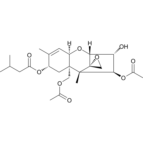
T-2 toxin is a trichothecene mycotoxin produced by fungi of the genus Fusarium. It is a common contaminant in food and feedstuffs of cereal origin and is known to cause a range of toxic effects in humans and animals. It has a role as a mycotoxin, a cardiotoxic agent, a neurotoxin, an environmental contaminant, an apoptosis inducer, a DNA synthesis inhibitor and a fungal metabolite. It is a trichothecene, an acetate ester and an organic heterotetracyclic compound. It is functionally related to a HT-2 toxin.
T2 Toxin has been reported in Fusarium heterosporum, Fusarium chlamydosporum, and other organisms with data available. T-2 Toxin is a type A trichothecene mycotoxin produced by Fusarium langsethiae, Fusarium poae, and Fusarium sporotrichioides that interferes with the metabolism of membrane phospholipids by inhibiting protein synthesis and disrupting DNA and RNA synthesis. It is frequently responsible for the contamination of various grain crops and elicits a severe inflammatory reaction in animals. A potent mycotoxin produced in feedstuffs by several species of the genus FUSARIUM. It elicits a severe inflammatory reaction in animals and has teratogenic effects. Mechanism of Action Studies with whole cells, cell-free protein synthetic system, & acid-insol cell fractions of Tetrahymena pyriformis indicated that T-2 toxin inhibited protein synthesis by impairing the 60 S ribosome subunit & inhibited RNA & dna synthesis by disturbing the cell membrane function. T2-trichothecene binds in vitro to active SH groups of creatine phosphokinase, lactate dehydrogenase & alcohol dehydrogenase, inhibiting their catalytic activity. The high affinity of T2-trichothecene & higher trichothecenes to SH compounds provides a molecular basis for an interaction with the spindle fiber mechanism. ... T-2 toxin could inhibit synthesis of DNA and RNA both in vivo (0.75 mg/kg bw single or multiple doses) and in vitro (> 0.1-1 ng/mL). For more Mechanism of Action (Complete) data for T-2 TOXIN (14 total), please visit the HSDB record page. |
| 分子式 |
C24H34O9
|
|---|---|
| 分子量 |
466.52136
|
| 精确质量 |
466.22
|
| 元素分析 |
C, 61.79; H, 7.35; O, 30.86
|
| CAS号 |
21259-20-1
|
| 相关CAS号 |
T-2 Toxin-13C24
|
| PubChem CID |
5284461
|
| 外观&性状 |
White to off-white solid powder
|
| 密度 |
1.3±0.1 g/cm3
|
| 沸点 |
544.9±50.0 °C at 760 mmHg
|
| 熔点 |
151.5℃
|
| 闪点 |
177.0±23.6 °C
|
| 蒸汽压 |
0.0±3.3 mmHg at 25°C
|
| 折射率 |
1.547
|
| LogP |
2.25
|
| tPSA |
120.89
|
| 氢键供体(HBD)数目 |
1
|
| 氢键受体(HBA)数目 |
9
|
| 可旋转键数目(RBC) |
9
|
| 重原子数目 |
33
|
| 分子复杂度/Complexity |
881
|
| 定义原子立体中心数目 |
8
|
| SMILES |
CC1=C[C@@H]2[C@](C[C@@H]1OC(=O)CC(C)C)([C@]3([C@@H]([C@H]([C@H]([C@@]34CO4)O2)O)OC(=O)C)C)COC(=O)C
|
| InChi Key |
BXFOFFBJRFZBQZ-QYWOHJEZSA-N
|
| InChi Code |
InChI=1S/C24H34O9/c1-12(2)7-18(27)32-16-9-23(10-29-14(4)25)17(8-13(16)3)33-21-19(28)20(31-15(5)26)22(23,6)24(21)11-30-24/h8,12,16-17,19-21,28H,7,9-11H2,1-6H3/t16-,17+,19+,20+,21+,22+,23+,24-/m0/s1
|
| 化学名 |
[(1S,2R,4S,7R,9R,10R,11S,12S)-11-acetyloxy-2-(acetyloxymethyl)-10-hydroxy-1,5-dimethylspiro[8-oxatricyclo[7.2.1.02,7]dodec-5-ene-12,2'-oxirane]-4-yl] 3-methylbutanoate
|
| 别名 |
T-2 TOXIN; T2 Toxin; 21259-20-1; T2-Trichothecene; Mycotoxin T-2; Isaritoxin; Fusariotoxine T2; Fusariotoxin T-2;
|
| HS Tariff Code |
2934.99.9001
|
| 存储方式 |
Powder -20°C 3 years 4°C 2 years In solvent -80°C 6 months -20°C 1 month |
| 运输条件 |
Room temperature (This product is stable at ambient temperature for a few days during ordinary shipping and time spent in Customs)
|
| 溶解度 (体外实验) |
DMSO : ≥ 100 mg/mL (~214.35 mM)
|
|---|---|
| 溶解度 (体内实验) |
配方 1 中的溶解度: ≥ 2.5 mg/mL (5.36 mM) (饱和度未知) in 10% DMSO + 40% PEG300 + 5% Tween80 + 45% Saline (这些助溶剂从左到右依次添加,逐一添加), 澄清溶液。
例如,若需制备1 mL的工作液,可将100 μL 25.0 mg/mL澄清DMSO储备液加入到400 μL PEG300中,混匀;然后向上述溶液中加入50 μL Tween-80,混匀;加入450 μL生理盐水定容至1 mL。 *生理盐水的制备:将 0.9 g 氯化钠溶解在 100 mL ddH₂O中,得到澄清溶液。 配方 2 中的溶解度: ≥ 2.5 mg/mL (5.36 mM) (饱和度未知) in 10% DMSO + 90% (20% SBE-β-CD in Saline) (这些助溶剂从左到右依次添加,逐一添加), 澄清溶液。 例如,若需制备1 mL的工作液,可将 100 μL 25.0 mg/mL澄清DMSO储备液加入900 μL 20% SBE-β-CD生理盐水溶液中,混匀。 *20% SBE-β-CD 生理盐水溶液的制备(4°C,1 周):将 2 g SBE-β-CD 溶解于 10 mL 生理盐水中,得到澄清溶液。 View More
配方 3 中的溶解度: ≥ 2.5 mg/mL (5.36 mM) (饱和度未知) in 10% DMSO + 90% Corn Oil (这些助溶剂从左到右依次添加,逐一添加), 澄清溶液。 1、请先配制澄清的储备液(如:用DMSO配置50 或 100 mg/mL母液(储备液)); 2、取适量母液,按从左到右的顺序依次添加助溶剂,澄清后再加入下一助溶剂。以 下列配方为例说明 (注意此配方只用于说明,并不一定代表此产品 的实际溶解配方): 10% DMSO → 40% PEG300 → 5% Tween-80 → 45% ddH2O (或 saline); 假设最终工作液的体积为 1 mL, 浓度为5 mg/mL: 取 100 μL 50 mg/mL 的澄清 DMSO 储备液加到 400 μL PEG300 中,混合均匀/澄清;向上述体系中加入50 μL Tween-80,混合均匀/澄清;然后继续加入450 μL ddH2O (或 saline)定容至 1 mL; 3、溶剂前显示的百分比是指该溶剂在最终溶液/工作液中的体积所占比例; 4、 如产品在配制过程中出现沉淀/析出,可通过加热(≤50℃)或超声的方式助溶; 5、为保证最佳实验结果,工作液请现配现用! 6、如不确定怎么将母液配置成体内动物实验的工作液,请查看说明书或联系我们; 7、 以上所有助溶剂都可在 Invivochem.cn网站购买。 |
| 制备储备液 | 1 mg | 5 mg | 10 mg | |
| 1 mM | 2.1435 mL | 10.7177 mL | 21.4353 mL | |
| 5 mM | 0.4287 mL | 2.1435 mL | 4.2871 mL | |
| 10 mM | 0.2144 mL | 1.0718 mL | 2.1435 mL |
1、根据实验需要选择合适的溶剂配制储备液 (母液):对于大多数产品,InvivoChem推荐用DMSO配置母液 (比如:5、10、20mM或者10、20、50 mg/mL浓度),个别水溶性高的产品可直接溶于水。产品在DMSO 、水或其他溶剂中的具体溶解度详见上”溶解度 (体外)”部分;
2、如果您找不到您想要的溶解度信息,或者很难将产品溶解在溶液中,请联系我们;
3、建议使用下列计算器进行相关计算(摩尔浓度计算器、稀释计算器、分子量计算器、重组计算器等);
4、母液配好之后,将其分装到常规用量,并储存在-20°C或-80°C,尽量减少反复冻融循环。
计算结果:
工作液浓度: mg/mL;
DMSO母液配制方法: mg 药物溶于 μL DMSO溶液(母液浓度 mg/mL)。如该浓度超过该批次药物DMSO溶解度,请首先与我们联系。
体内配方配制方法:取 μL DMSO母液,加入 μL PEG300,混匀澄清后加入μL Tween 80,混匀澄清后加入 μL ddH2O,混匀澄清。
(1) 请确保溶液澄清之后,再加入下一种溶剂 (助溶剂) 。可利用涡旋、超声或水浴加热等方法助溶;
(2) 一定要按顺序加入溶剂 (助溶剂) 。