
| 规格 | 价格 | 库存 | 数量 |
|---|---|---|---|
| 1mg |
|
||
| 5mg |
|
||
| 10mg |
|
||
| 50mg |
|
||
| 100mg |
|
||
| Other Sizes |
|
| 靶点 |
S1PR1 1.03 nM (EC50) S1PR5 8.6 nM (EC50)
|
|---|---|
| 体外研究 (In Vitro) |
Ozanimod (RPC-1063) 盐酸盐作为具有 [35S]-GTPgS 结合的跨物种 S1P5 的 S1P 受体调节剂,表现出内在活性和效力。人 S1P1、食蟹猴 S1P1、小鼠 S1P1、大鼠 S1P1 和犬 S1P1 的 EC50 值分别为 1.03 nM、1.29 nM、0.90 nM、1.02 nM 和 0.61 nM;食蟹猴 S1P5、小鼠 S1P5、大鼠 S1P5 和犬 S1P5 的浓度分别为 8.6 nM、15.9 nM、957.5 nM、2032.7 nM 和 1662.0 nM[1]。通过改变小鼠序列中的丙氨酸,奥扎莫德盐酸盐将 EC50 的效力从 mS1P5 的 958 nM 恢复到 mS1P5_A120T 的 6.7 nM,几乎与 hS1P5 的 EC50 8.6 nM 一致[1]。 ozanimod 盐酸盐对 hS1P5、mS1P5 和 mS1P5 _A120T 的结合亲和力分别为 2.0 nM、59.9 nM 和 5.6 nM[1]。除了 [3H]-A971432 与 S1P5D 的饱和结合值为 8.75 nM 外,ozanimod 盐酸盐还具有 [3H]-ozanimod 与 hS1P5 和 mS1P5_A120T 的饱和结合,KD 值分别为 6.56 nM 和 7.35 nM[1]。
|
| 体内研究 (In Vivo) |
奥扎莫德 (RPC-1063) 盐酸盐(口服强饲;0.05、0.2 或 1 mg/kg;每天一次;连续 14 天)暴露足以参与 S1P1 的结果是循环淋巴细胞减少、疾病评分和体重减轻,但不是S1P5。此外,脊髓炎症、脱髓鞘和凋亡细胞的水平降低,神经丝光的循环水平也降低,神经丝光是神经元变性的标志[1]。在毒素攻击期间,盐酸奥扎莫德(口服强饲;5 mg/kg;每日一次)减少髓鞘质损失和轴突降解,但它不会促进中毒后髓鞘再生的增加[1]。口服奥扎莫德盐酸盐(1 或 5 mg/kg,连续 7 天)治疗的小鼠表现出优异的药代动力学[1]。
|
| 酶活实验 |
使用 LiveBLAzer-FRET B/G 测定通过细胞信号传导测定检测 cAMP (S1P1R) 或 β-arrestin (S1P4R) 信号传导。按照制造商的指示,在 384 孔板中进行三次重复测定。首先使用 20%(2-羟丙基)-β-环糊精按 1:10 稀释化合物原液,并在 -50°C 下在 100% DMSO 中保持 10 mM。每 40 倍最终测定浓度(10 mM),就会生成 10 点剂量反应曲线。 Hepes 7.4 pH,含 0.1% Pluronic F-127。将 80 μM 毛喉素添加到 S1P1R 测定的稀释液中。简而言之,每孔 104 个细胞在 37°C 下培养 4 小时,可获得配体的剂量反应。添加丙磺舒和 CC4-AM 底物,并在 37°C 下再孵育两小时后,使用 Spectramax M5 检查样品。 S1P1R cAMP 测定的数据标准化为 2 μM 毛喉素产生的最高荧光。在 GTPγS 结合测定中,将 1–5 μg/孔膜蛋白在 96 孔板中与 10 μM GDP、100–500 μg/孔麦芽凝集素 PVT SPA 珠在 50 mM HEPES、100 mM NaCl 中孵育 15 分钟, 10 mM MgCl2、20 μg/ml 皂苷和 0.1% 不含脂肪酸 BSA。添加化合物和200 pM GTP [35S] 1250Ci/mmol后,将板孵育120分钟,然后在300g下离心5分钟。 TopCount 仪器用于检测放射性。称为斜率的四参数变量用于拟合所有数据。使用 TopCount 仪器检测放射性。 GraphPad Prism 的四参数变量斜率非线性回归用于拟合所有数据,以确定与 S1P 相关的最大功效和半最大有效浓度 (EC50)。
|
| 细胞实验 |
Ozanimod (RPC1063) 是 S1P1 和 S1P5 受体的特殊激动剂。 S1P1 受体抑制 cAMP 生成和 [35S]-GTPγS 结合,EC50 值为 160 ± 60 pM 和 410 ±分别为下午 160 点。与S1P5受体相关,奥扎莫德的83% Emax值为11±4.3nM。孵育一小时后,RPC1063几乎完全且持续地减少S1P1受体-HEK293T细胞表面的S1P1受体再表达。这是在浓度大于 10 nM 时观察到的。
|
| 动物实验 |
Animal/Disease Models: Experimental Autoimmune Encephalomyelitis Model[1]
Doses: 0.05, 0.2, or 1 mg/kg Route of Administration: oral gavage; 0.05, 0.2, or 1 mg/kg; one time/day; for 14 days Experimental Results: Attenuated body weight loss, terminal disease scores were Dramatically attenuated with the 0.2 and 1 mg/kg doses and ALCs were Dramatically decreased in all dose groups. decreased spinal cord inflammation and demyelination, as well as attenuated the number of spinal cord apoptotic cells, and Dramatically decreased the levels of circulating neurofilament light at the top dose of 1 mg/kg. Animal/Disease Models: Cuprizone/Rapamycin Demyelination Model[1] Doses: 5 mg/kg Route of Administration: oral gavage; 5 mg/kg; once-daily Experimental Results: Protected neuronal axons, preventing breakage and ovoid formation in the corpus callosum of CPZ/Rapa treated mice. Dramatically attenuated the extent to which the corpus callosum demonstrated decreased myelin content as visualized by MRI. Did not result in enhanced myelin content. MOG35–55 Experimental Autoimmune Encephalomyelitis Model[1] Experimental autoimmune encephalomyelitis (EAE) was induced in 10-week-old female C57BL/6 mice (Taconic Biosciences, Rensselaer, NY) by subcutaneous immunization with an emulsion of myelin oligodendrocyte glycoprotein 35–55 (MOG35–55) in complete Freund’s adjuvant (CFA) followed by intraperitoneal injections of pertussis toxin 2 and 24 hours later. Mice received two subcutaneous injections, one in the upper and one in the lower back, of 0.1 ml MOG35–55/CFA emulsion per site, and both intraperitoneal injections of pertussis toxin were 100 ng per dose at a volume of 0.1 ml per dose. The study was performed at Hooke Laboratories (Lawrence, MA) using Hooke Kit MOG35–55/CFA Emulsion PTX number EK-2110. Female mice were selected for EAE experimentation since more females than males suffer clinically with MS as well as other autoimmune diseases (Voskuhl, 2011). EAE is an immune-driven preclinical model of MS, and female mice are reported to experience greater severity of disease (Papenfuss et al., 2004; Rahn et al., 2014). Mice were assessed daily and upon the first emergence of signs of disease, randomized into treatment groups (n = 12) on the basis of comparable group average values for time of EAE onset and disease score at the onset of treatment. Dosing was initiated on the first day of EAE disease via once daily oral gavage of vehicle (5% v/v DMSO, 5% v/v Tween20, 90% v/v Milli-Q water; 5 ml/kg) or ozanimod at doses of 0.05, 0.2, or 1 mg/kg for 14 consecutive days. Efficacy was evaluated by recording daily visual EAE disease scores, as described previously by Scott et al. (2016), as well as body weight measurement three times per week. Approximately 24 hours after the final dose, a blood sample was collected in EDTA coagulant for the assessment of absolute numbers of circulating lymphocytes by differential count, and a separate plasma sample was processed and stored at −80°C for subsequent analysis of neurofilament light by Quanterix (Lexington, MA) using the Simoa NF-light Advantage kit (102258). Mice were anesthetized and perfused with phosphate buffered saline, and the spinal cords collected and stored in 10% buffered formalin for imaging analysis. For each mouse spinal cord, three hematoxylin and eosin sections were prepared and analyzed for the number of inflammatory foci (approximately 20 cells per foci), estimation of demyelinated area (scores of 0–5 representing <5%, 5 to 20%, 20 to 40%, 40 to 60%, 60 to 80%, and 80 to 100% demyelinated area, respectively, and as defined by interruption of normal structure such as pallor and vacuolation consistent with edema and demyelination, as well as dilated axons) and apoptotic cell counts. Histologic analysis was performed by a pathologist blinded to the experimental design and readouts. Cuprizone/Rapamycin Demyelination Model: Neuroprotection and Remyelination[1] Cuprizone/rapamycin-induced demyelination was initiated in 8-week-old male C57BL/6J mice (Jackson Laboratories, Bar Harbor, ME) by ad libitum access to normal rodent diet (Harlan Teklad, Madison, WI) containing cuprizone (0.3% w/w) for a period of 6 weeks with once daily intraperitoneal injection of rapamycin. Rapamycin was prepared fresh daily at 10 mg/kg at a volume of 5 ml/kg in 5% v/v pure ethanol/5% v/v Tween 80/5% PEG1000, aqueous. Age-matched control mice had ad libitum access to the same diet not containing cuprizone and received daily intraperitoneal injection with vehicle. Mice were group housed 4 to 5 per cage, and fresh food was provided three times weekly. All mice had ad libitum access to reverse osmosis filtered, acidified palatable drinking water at a pH level of 2.5 to 3.0. The study was performed at Renovo Neural, Inc. (Cleveland, OH). Male mice were chosen for the demyelination model since a number of studies have reported that females are more resistant to the toxin and hence more robust demyelination is observed in males (MacArthur and Papanikolaou, 2014). View More
After 2 weeks of acclimation, mice were randomly assigned to dose groups and received once-daily oral gavage administration of vehicle (5% v/v DMSO, 5% v/v Tween20, 90% v/v Milli-Q water; 5 ml/kg) or ozanimod 5 mg/kg after the dosing and sample collection/testing regimen depicted in Fig. 1. For the assessment of ozanimod on neuroprotection and demyelination, dosing was initiated on day 1 concurrent with cuprizone/rapamycin and continued daily for 6 weeks. For the assessment of ozanimod’s effect on remyelination, daily dosing was also initiated on day 1 but continued beyond the 6-week cuprizone/rapamycin challenge for a further 12-week period (weeks 7–18 of the study). Mice in the remyelination arms of assessment were discontinued from cuprizone diet and daily intraperitoneal rapamycin injection at the end of the 6-week challenge period and returned to normal rodent diet. Pharmacokinetics[1] The pharmacokinetic profiles of ozanimod and its primary active rodent metabolite, RP101075, are similar in male and female C57BL/6J mice and so were assessed in plasma and brains of 8-week-old male C57BL/6J mice (Jackson Laboratories) after daily oral dosing with ozanimod for 7 consecutive days. Ozanimod was dosed at either 1 or 5 mg/kg in the same vehicle as used for the MOG35–55 EAE and cuprizone/rapamycin in vivo efficacy studies and terminal plasma, and brain samples were collected 3, 6, and 24 hours after the seventh daily dose of ozanimod. Of note, in the clinical setting, the dosing of ozanimod involves a dose titration to avoid and potential risk of mechanism-based bradycardia, but this is not adopted when assessing efficacy in preclinical studies where dosing is initiated straight away with the dose to be assessed without titration. Brains were homogenized in acetonitrile at a 1:3 (w/v) ratio using a Biospec Bead Beater-16 with 1 mm glass beads and proteins precipitated further with a 1:10 dilution in acetonitrile to 1:30 (w/v) final. Plasma proteins were precipitated with acetonitrile at a 1:3 ratio (v/v). Samples were centrifuged and supernatants were analyzed by liquid chromatography–mass spectrometry (LC-MS/MS). For the tissue analysis, a standard curve was prepared using homogenized brain samples from untreated animals. A 10-point standard curve of ozanimod or RP101075 spanning a range of 0.046 nM to 500 nM was included with each bioanalytical run using a Kinetex C18 2.6μ 30 × 3 mm column (Phenomenex Inc., Torrance, CA), 0.1% formic acid in deionized H2O mobile phase A, and 0.1% formic acid in acetonitrile mobile phase B. Data were collected and analyzed using Analyst software version 1.5.1. |
| 毒性/毒理 (Toxicokinetics/TK) |
Effects During Pregnancy and Lactation
◉ Summary of Use during Lactation Although ozanimod and its active metabolites are highly bound in maternal plasma and unlikely to reach the breastmilk in large amounts, it is potentially toxic to the breastfed infant. Because there is no published experience with ozanimod during breastfeeding, expert opinion generally recommends that the closely related drug fingolimod should be avoided during breastfeeding, especially while nursing a newborn or preterm infant. Some guidelines recommend avoiding ozanimod during breastfeeding because of a lack of data; however, the manufacturer's labeling does not recommend against the use of ozanimod in breastfeeding. ◉ Effects in Breastfed Infants Relevant published information was not found as of the revision date. ◉ Effects on Lactation and Breastmilk Relevant published information was not found as of the revision date. |
| 参考文献 | |
| 其他信息 |
Ozanimod Hydrochloride is the hydrochloride salt form of ozanimod, an orally bioavailable sphingosine-1-phosphate (S1P) receptors 1 (S1PR1, S1P1) and 5 (S1PR5, S1P5) modulator, with potential anti-inflammatory and immunomodulating activities. Upon oral administration, ozanimod selectively targets and binds to S1PR1 on lymphocytes and induces S1PR1 internalization and degradation. This results in the sequestration of lymphocytes in lymph nodes. By preventing egress of lymphocytes, ozanimod reduces both the amount of circulating peripheral lymphocytes and the infiltration of lymphocytes into target tissues. This prevents a lymphocyte-mediated immune response and may reduce inflammation. S1PR1, a G-protein coupled receptor, plays a key role in lymphocyte migration from lymphoid tissues. Modulation of S1PR5 by ozanimod may be neuroprotective.
See also: Ozanimod (has active moiety). Drug Indication Multiple sclerosisZeposia is indicated for the treatment of adult patients with relapsing remitting multiple sclerosis (RRMS) with active disease as defined by clinical or imaging features. Ulcerative colitisZeposia is indicated for the treatment of adult patients with moderately to severely active ulcerative colitis (UC) who have had an inadequate response, lost response, or were intolerant to either conventional therapy or a biologic agent. Treatment of multiple sclerosis Treatment of Crohn's disease Treatment of ulcerative colitis |
| 分子式 |
C23H25CLN4O3
|
|---|---|
| 分子量 |
440.92
|
| 精确质量 |
440.161
|
| 元素分析 |
C, 62.65; H, 5.72; Cl, 8.04; N, 12.71; O, 10.89
|
| CAS号 |
1618636-37-5
|
| 相关CAS号 |
Ozanimod;1306760-87-1
|
| PubChem CID |
91618104
|
| 外观&性状 |
White to off-white solid powder
|
| tPSA |
104Ų
|
| 氢键供体(HBD)数目 |
3
|
| 氢键受体(HBA)数目 |
7
|
| 可旋转键数目(RBC) |
7
|
| 重原子数目 |
31
|
| 分子复杂度/Complexity |
609
|
| 定义原子立体中心数目 |
1
|
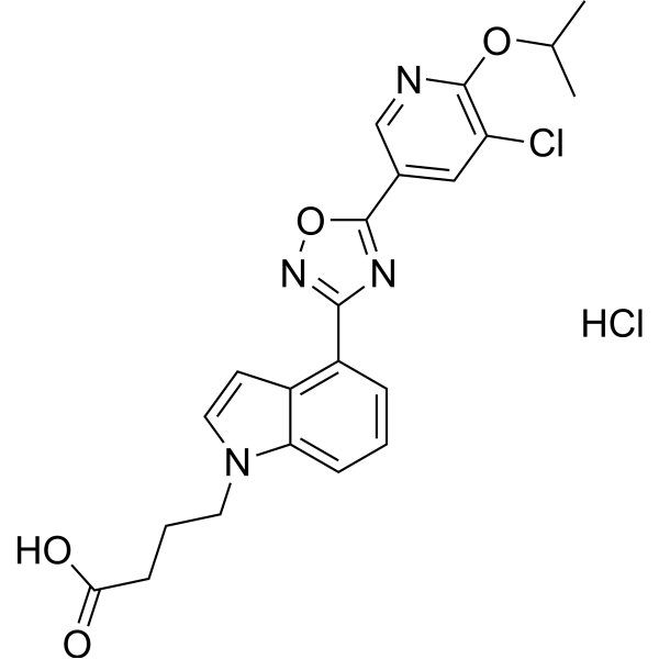
| SMILES |
Cl[H].O1C(C2C([H])=C([H])C(=C(C#N)C=2[H])OC([H])(C([H])([H])[H])C([H])([H])[H])=NC(C2C([H])=C([H])C([H])=C3C=2C([H])([H])C([H])([H])[C@]3([H])N([H])C([H])([H])C([H])([H])O[H])=N1
|
| InChi Key |
KBXLMOYQNDMHQT-QGZVFWFLSA-N
|
| InChi Code |
InChI=1S/C23H24N4O3.ClH/c1-14(2)29-21-9-6-15(12-16(21)13-24)23-26-22(27-30-23)19-5-3-4-18-17(19)7-8-20(18)25-10-11-28;/h3-6,9,12,14,20,25,28H,7-8,10-11H2,1-2H3;1H/t20-;/m0./s1
|
| 化学名 |
(S)-5-(3-(1-((2-hydroxyethyl)amino)-2,3-dihydro-1H-inden-4-yl)-1,2,4-oxadiazol-5-yl)-2-isopropoxybenzonitrile hydrochloride
|
| 别名 |
RPC-1063 HCl; RPC1063 hydrochloride; RPC-1063 hydrochloride; Zeposia; UNII-3UPR33JAAM; 3UPR33JAAM
|
| HS Tariff Code |
2934.99.9001
|
| 存储方式 |
Powder -20°C 3 years 4°C 2 years In solvent -80°C 6 months -20°C 1 month 注意: 请将本产品存放在密封且受保护的环境中,避免吸湿/受潮。 |
| 运输条件 |
Room temperature (This product is stable at ambient temperature for a few days during ordinary shipping and time spent in Customs)
|
| 溶解度 (体外实验) |
DMSO: 200 mg/mL (453.60 mM)
|
|---|---|
| 溶解度 (体内实验) |
配方 1 中的溶解度: ≥ 5 mg/mL (11.34 mM) (饱和度未知) in 10% DMSO + 40% PEG300 + 5% Tween80 + 45% Saline (这些助溶剂从左到右依次添加,逐一添加), 澄清溶液。
例如,若需制备1 mL的工作液,可将100 μL 50.0 mg/mL澄清DMSO储备液加入400 μL PEG300中,混匀;然后向上述溶液中加入50 μL Tween-80,混匀;加入450 μL生理盐水定容至1 mL。 *生理盐水的制备:将 0.9 g 氯化钠溶解在 100 mL ddH₂O中,得到澄清溶液。 配方 2 中的溶解度: ≥ 5 mg/mL (11.34 mM) (饱和度未知) in 10% DMSO + 90% (20% SBE-β-CD in Saline) (这些助溶剂从左到右依次添加,逐一添加), 澄清溶液。 例如,若需制备1 mL的工作液,可将 100 μL 50.0 mg/mL澄清DMSO储备液加入900 μL 20% SBE-β-CD生理盐水溶液中,混匀。 *20% SBE-β-CD 生理盐水溶液的制备(4°C,1 周):将 2 g SBE-β-CD 溶解于 10 mL 生理盐水中,得到澄清溶液。 View More
配方 3 中的溶解度: ≥ 5 mg/mL (11.34 mM) (饱和度未知) in 10% DMSO + 90% Corn Oil (这些助溶剂从左到右依次添加,逐一添加), 澄清溶液。 1、请先配制澄清的储备液(如:用DMSO配置50 或 100 mg/mL母液(储备液)); 2、取适量母液,按从左到右的顺序依次添加助溶剂,澄清后再加入下一助溶剂。以 下列配方为例说明 (注意此配方只用于说明,并不一定代表此产品 的实际溶解配方): 10% DMSO → 40% PEG300 → 5% Tween-80 → 45% ddH2O (或 saline); 假设最终工作液的体积为 1 mL, 浓度为5 mg/mL: 取 100 μL 50 mg/mL 的澄清 DMSO 储备液加到 400 μL PEG300 中,混合均匀/澄清;向上述体系中加入50 μL Tween-80,混合均匀/澄清;然后继续加入450 μL ddH2O (或 saline)定容至 1 mL; 3、溶剂前显示的百分比是指该溶剂在最终溶液/工作液中的体积所占比例; 4、 如产品在配制过程中出现沉淀/析出,可通过加热(≤50℃)或超声的方式助溶; 5、为保证最佳实验结果,工作液请现配现用! 6、如不确定怎么将母液配置成体内动物实验的工作液,请查看说明书或联系我们; 7、 以上所有助溶剂都可在 Invivochem.cn网站购买。 |
| 制备储备液 | 1 mg | 5 mg | 10 mg | |
| 1 mM | 2.2680 mL | 11.3399 mL | 22.6799 mL | |
| 5 mM | 0.4536 mL | 2.2680 mL | 4.5360 mL | |
| 10 mM | 0.2268 mL | 1.1340 mL | 2.2680 mL |
1、根据实验需要选择合适的溶剂配制储备液 (母液):对于大多数产品,InvivoChem推荐用DMSO配置母液 (比如:5、10、20mM或者10、20、50 mg/mL浓度),个别水溶性高的产品可直接溶于水。产品在DMSO 、水或其他溶剂中的具体溶解度详见上”溶解度 (体外)”部分;
2、如果您找不到您想要的溶解度信息,或者很难将产品溶解在溶液中,请联系我们;
3、建议使用下列计算器进行相关计算(摩尔浓度计算器、稀释计算器、分子量计算器、重组计算器等);
4、母液配好之后,将其分装到常规用量,并储存在-20°C或-80°C,尽量减少反复冻融循环。
计算结果:
工作液浓度: mg/mL;
DMSO母液配制方法: mg 药物溶于 μL DMSO溶液(母液浓度 mg/mL)。如该浓度超过该批次药物DMSO溶解度,请首先与我们联系。
体内配方配制方法:取 μL DMSO母液,加入 μL PEG300,混匀澄清后加入μL Tween 80,混匀澄清后加入 μL ddH2O,混匀澄清。
(1) 请确保溶液澄清之后,再加入下一种溶剂 (助溶剂) 。可利用涡旋、超声或水浴加热等方法助溶;
(2) 一定要按顺序加入溶剂 (助溶剂) 。
 Spns2-IN-2
 GSK2018682 hydrochloride
 BMS-986104
 SLF80821178 hydrochloride
InvivoChem的所有产品仅用于作科学研究,不面向患者销售
Copyright 2020 InvivoChem LLC | All Rights Reserved 粤ICP备20063088号-1


