
| 规格 | 价格 | 库存 | 数量 |
|---|---|---|---|
| 2mg |
|
||
| 5mg |
|
||
| 10mg |
|
||
| 25mg |
|
||
| 50mg |
|
||
| Other Sizes |
|

| 靶点 |
α7 nAChR/α7 nicotinic acetylcholine receptor
|
|---|---|
| 体外研究 (In Vitro) |
在大鼠脑匀浆中,PNU-282987(化合物 C7)的 Ki 为 27 nM,可替代 R7 选择性拮抗剂甲基乌头碱 (MLA) [1]。 PNU-282987 的 EC50 为 154 nM,具有 α7 nAChR 激动剂活性 [1]。此外,PNU-282987 还能阻断 5-HT3 受体,IC50 为 4541nM[1]。
使用了几种试验来验证嵌合体试验可用于鉴定天然受体的激动剂。在结合试验中,PNU-282987以27 nM的Ki从大鼠脑匀浆中置换了α7选择性拮抗剂甲基乌头碱(MLA)。此外,当应用于培养的大鼠海马神经元时,PNU-282987诱发了一种快速脱敏的内向全细胞电流,该电流具有浓度依赖性,可被MLA阻断,与α7受体的开放一致[1] 还评估了PNU-282987对相关受体的选择性。研究人员特别关注神经肌肉接头形式的受体(α1β1γδ)和主要神经节nAChR(α3β4)的激动作用。这些受体的激活被证明会导致非特异性激动剂如依巴替丁和尼古丁的许多不良反应。15 PNU-282987在两种受体亚型上均未检测到高达100μM的激动剂活性,拮抗剂活性可忽略不计(IC50≥60μM)。此外,PNU-282987在1μM下没有显著取代大鼠脑匀浆中的氚化金雀花碱(14%的抑制率),表明其对α4β2亚型具有高选择性。16关于5-HT3受体,PNU-282987以1662 nM的Ki取代了氚化的GR-65630,17与5-HT3R的高选择性1(超过500倍)相比,α7的选择性约为62倍。在基于细胞的FLIPR测定中,发现PNU-282987是5-HT3受体的功能性拮抗剂(IC50=4541nM)。在Cerep(法国Rueil-Malmaison)的32个受体、离子通道和酶的筛选中评估了PNU-282987的更广泛选择性。在1μM的测试浓度下,PNU-282987对除5-HT3受体外的所有靶点的特异性结合或酶活性产生了<30%的抑制[1]。 |
| 体内研究 (In Vivo) |
PNU-282987(化合物 C7)(iv;1、3 mg/kg)可以逆转门控缺陷[1]。在大鼠海马神经元中,PNU-282987 (30 μM) 以浓度依赖性和 MLA 可阻断的方式刺激电流 [1]。
研究人员还在感觉门控受损的大鼠模型中测试了PNU-282987,该模型已用已知的α7部分激动剂GTS-21进行了验证。18全身给药d-苯丙胺(0.3或1mg/kg,iv)显著扰乱了麻醉大鼠的听觉门控,因为条件反射同时降低,测试反应相应增加。随后给予α7 nAChR激动剂PNU-282987(iv,1或3 mg/kg,n=10)显著逆转了苯丙胺诱导的门控缺陷(图4)。相比之下,在对照组大鼠中应用赋形剂并没有使苯丙胺诱导的门控缺陷正常化(n=9)。此外,PNU-282987(1mg/kg)对麻醉大鼠的正常门控(n=4)没有显著影响。 |
| 酶活实验 |
脑匀浆结合试验([3H]-MLA,[3H]-金雀花碱,[3H]-GR65630):[1]
通过断头处死雄性Sprague-Dawley大鼠(300-350g),快速解剖大脑(全脑减去小脑),称重并在50℃下使用旋转杵在9体积/g湿重的0.32M冰冷蔗糖中均质化(10次上下击打)。将匀浆在40°C下以1000 x g离心10分钟。收集上清液,在40°C.下以20000 x g离心20分钟。将所得沉淀重新悬浮至蛋白质浓度为1-8mg/ml。将5ml匀浆的等分试样在-80°C.下冷冻,直至需要进行分析。在测定当天,将等分试样在室温下解冻,并用含有4.16 mM NaHCO3、0.44 mM KH2PO4、127 mM NaCl、5.36 mM KCl、1.26 mM CaCl2和0.98 mM MgCl2的Kreb's-20 mM HEPES缓冲液pH 7.0(在室温下)稀释,从而每个试管中添加25-150 mg蛋白质。以牛血清白蛋白为标准,采用Bradford法测定蛋白质浓度。对于α7,在放射性配体之前添加1µM MLA的情况下平行孵育的组织中测定了非特异性结合,在竞争研究中,在添加约3 nM[3H]-MLA(25 Ci/mmol)之前,将化合物以越来越高的浓度添加到试管中。对于α4,在放射性配体之前添加1 mM(-)-尼古丁的情况下平行孵育的组织中测定了非特异性结合,在竞争研究中,在添加约1.0 nM[3H]-金雀花碱之前,将化合物以越来越高的浓度添加到试管中。对于5-HT3,在放射性配体之前添加1µM ICS-205930的情况下,在平行孵育的组织中测定非特异性结合,在竞争研究中,在添加约0.45 nM[3H]-GR65630之前,将化合物以越来越高的浓度添加到试管中。对于所有结合试验,将0.4 ml匀浆加入含有缓冲液、试验化合物和放射性配体的试管中,并在25°下以0.5 ml的最终体积孵育1小时。通过安装在48孔Brandel细胞采集器上的Whatman GF/B玻璃滤纸进行快速真空过滤,终止培养。过滤器预先浸泡在50 mM Tris-HCl pH 7.0-0.05%聚乙烯亚胺中。用5ml等分的0.9%冷盐水洗涤过滤器两次,然后通过液体闪烁光谱法计算放射性。抑制常数(Ki)是通过将数据拟合到Cheng-Prusoff方程中获得的放射性配体结合的浓度依赖性抑制来计算的。PNU-282987在该测定中的Ki为27±1nM(n=48)
PNU-282987在1µM时没有显著置换大鼠脑匀浆中的氚化金雀花碱(抑制率=14±4%,n=13)。就5-HT3受体而言,PNU-28298.7置换了氚化GR-65630,Ki为1662±331 nM(n=10)
|
| 动物实验 |
Patch-clamp electrophysiology: Cultured neurons were prepared according to Brewer . Briefly, Sprague- Dawley rats (postnatal day 3) were killed by decapitation and their brains were removed and placed in ice cold Hibernate-A medium. Hippocampal regions were gently removed, cut into small pieces and placed in Hibernate-A medium with 1 mg/ml papain for 60 min at 35°C. After digestion, the tissues were washed several times in Hibernate-A media and transferred to a 50 ml conical tube containing 6 ml Hibernate-A medium with 2% B-27 supplement. Neurons were dissociated by gentle trituration and plated onto poly-D-lysine/laminin coated coverslips at a density of 300 – 700 cells/mm2 , and transferred to 24-well tissue culture plates containing warmed culture medium composed of Neurobasal-A medium, B-27 supplement (2%), L-glutamine (0.5 mM), 100 U/ml penicillin, 100 mg/ml streptomycin, and 0.25 mg/ml Fungizone. Cells were maintained in a humidified incubator at 37°C and 6% CO2 for 1 – 2 weeks. The medium was changed after 24 h and then approximately every three days thereafter. Patch pipettes were pulled from borosilicate capillary glass using a Flaming/Brown micropipette puller and filled with an internal pipette solution composed of (in mM): CsCH3SO3 (126), CsCl (10), NaCl (4), MgCl2 (1), CaCl2 (0.5), EGTA (5), HEPES (10), ATP-Mg (3), GTP-Na (0.3), phosphocreatin (4), pH 7.2. The resistances of the patch pipettes when filled with internal solution ranged between 3 – 6 M•. All experiments were conducted at room temperature. Cultured cells were continuously superfused with an external bath solution containing (in mM): NaCl (140), KCl (5), CaCl2 (2), MgCl2 (1), HEPES (10), glucose (10), bicuculline (0.01), CNQX (0.005), D-AP-5 (0.005) tetrodotoxin (0.0005), pH 7.4. Compounds were dissolved in water or DMSO and diluted into the external bath solution containing a final DMSO concentration of 0.1% and delivered via a multibarrel fast perfusion system. Whole-cell currents were recorded using an Axopatch 200B amplifier (Axon Instruments, Union City, CA). Analog signals were filtered at 1/5 the sampling frequency, digitized, stored, and measured using pCLAMP software (Axon Instruments). All data are reported as mean ± SEM. [1]
Auditory gating assay. Experiments were performed on Male Sprague-Dawley rats (weighing 250 to 300 gm) under chloral hydrate anesthesia (400 mg/kg, IP). The femoral artery and vein were cannulated for monitoring arterial blood pressure and administration of drugs or additional doses of anaesthetic, respectively. Unilateral hippocampal field potential (EEG) was recorded by a metal monopolar macroelectrode placed into the CA3 region (co-ordinates: 3.0 – 3.5 mm posterior from the bregma, 2.6 –3.0 mm lateral and 3.8 – 4.0 mm ventral; Paxinos and Watson, 19863 ). Field potentials were amplified, filtered (0.1 – 100 Hz), displayed and recorded for on-line and off-line analysis (Spike3). Quantitative EEG analysis was performed by means of Fast Fourier Transformation (Spike3). The auditory stimulus consisted of a pair of 10 ms, 5 KHz tone bursts with a 0.5 s delay between the first “conditioning” stimulus and second “test” stimulus. Auditory evoked responses were computed by averaging of responses to 50 pairs of stimuli presented with an interstimulus interval of 10 s. Percentage of gating was determined by the formula: (1 - test amplitude/conditioning amplitude) x 100. Amphetamine (D-amphetamine sulfate, 0.3-1 mg/kg, IV) was administered in order to disrupt sensory gating. Recordings of evoke potentials commenced 5 min after amphetamine administration, and only rats showing gating deficit exceeding 20 % were used for subsequent evaluation of α7 nAChR agonists or vehicle. Statistical significance was determined by means of two-tailed paired Student’s t-test. [1] |
| 参考文献 | |
| 其他信息 |
A library of benzamides was tested for alpha7 nicotinic acetylcholine receptor (nAChR) agonist activity using a chimeric receptor in a functional, cell-based, high-throughput assay. From this library, quinuclidine benzamides were found to have alpha7 nAChR agonist activity. The SAR diverged from the activity of this compound class verses the 5-HT(3) receptor, a structural homologue of the alpha7 nAChR. PNU-282987, the most potent compound from this series, was also shown to open native alpha7 nAChRs in cultured rat neurons and to reverse an amphetamine-induced gating deficit in rats.[1]
In conclusion, using parallel synthesis, we have explored the SAR of tertiary-amine-containing arylamides as α7 nAChR agonists. Of the five structurally different amines tested, only 3-aminoquinuclidine yielded active analogues. The R enantiomers were more active than the corresponding S enantiomers. On the benzamide portion, small substituents in the para position gave the most active analogues. The most potent analogue in this series was the 4-chlorobenzamide, PNU-282987. Several experiments confirm that the α7-5HT3 chimera assay is predictive of native α7 nAChR activity. PNU-282987 displaced MLA from rat brain homogenates with a Ki of 27 nM, and it evoked currents in rat hippocampal neurons in a concentration-dependent and MLA blockable manner. We also tested this compound in a rat model of impaired sensory gating. Treatment of gating impaired rats with PNU-282987 led to a reversal of the gating deficit. These results demonstrate that (R)-3-aminoquinuclidine arylamides such as PNU-282897 are a template for finding α7 nAChR agonists that may be useful for treating the cognitive and attentional deficits of schizophrenia.[1] |
| 分子式 |
C14H17CLN2O
|
|---|---|
| 分子量 |
264.750582456589
|
| 精确质量 |
264.102
|
| 元素分析 |
C, 63.51; H, 6.47; Cl, 13.39; N, 10.58; O, 6.04
|
| CAS号 |
737727-12-7
|
| 相关CAS号 |
PNU-282987;123464-89-1;PNU-282987 free base;711085-63-1;(S)-PNU-282987 hydrochloride;128311-08-0
|
| PubChem CID |
14456380
|
| 外观&性状 |
White to off-white solid powder
|
| 密度 |
1.3±0.1 g/cm3
|
| 沸点 |
431.5±30.0 °C at 760 mmHg
|
| 闪点 |
214.8±24.6 °C
|
| 蒸汽压 |
0.0±1.0 mmHg at 25°C
|
| 折射率 |
1.612
|
| LogP |
2.49
|
| tPSA |
32.3
|
| 氢键供体(HBD)数目 |
1
|
| 氢键受体(HBA)数目 |
2
|
| 可旋转键数目(RBC) |
2
|
| 重原子数目 |
18
|
| 分子复杂度/Complexity |
307
|
| 定义原子立体中心数目 |
1
|
| SMILES |
C1CN2CCC1[C@@H](C2)NC(=O)C3=CC=C(C=C3)Cl
|
| InChi Key |
WECKJONDRAUFDD-CYBMUJFWSA-N
|
| InChi Code |
InChI=1S/C14H17ClN2O/c15-12-3-1-11(2-4-12)14(18)16-13-9-17-7-5-10(13)6-8-17/h1-4,10,13H,5-9H2,(H,16,18)/t13-/m1/s1
|
| 化学名 |
N-[(3S)-1-azabicyclo[2.2.2]octan-3-yl]-4-chlorobenzamide
|
| 别名 |
PNU-282987 S enantiomer free base; 737727-12-7; PNU-282987 (S enantiomer free base); N-[(3S)-1-azabicyclo[2.2.2]octan-3-yl]-4-chlorobenzamide; PNU-282987 S-Enantiomer; SCHEMBL10436185;
|
| HS Tariff Code |
2934.99.9001
|
| 存储方式 |
Powder -20°C 3 years 4°C 2 years In solvent -80°C 6 months -20°C 1 month |
| 运输条件 |
Room temperature (This product is stable at ambient temperature for a few days during ordinary shipping and time spent in Customs)
|
| 溶解度 (体外实验) |
May dissolve in DMSO (in most cases), if not, try other solvents such as H2O, Ethanol, or DMF with a minute amount of products to avoid loss of samples
|
|---|---|
| 溶解度 (体内实验) |
注意: 如下所列的是一些常用的体内动物实验溶解配方,主要用于溶解难溶或不溶于水的产品(水溶度<1 mg/mL)。 建议您先取少量样品进行尝试,如该配方可行,再根据实验需求增加样品量。
注射用配方
注射用配方1: DMSO : Tween 80: Saline = 10 : 5 : 85 (如: 100 μL DMSO → 50 μL Tween 80 → 850 μL Saline)(IP/IV/IM/SC等) *生理盐水/Saline的制备:将0.9g氯化钠/NaCl溶解在100 mL ddH ₂ O中,得到澄清溶液。 注射用配方 2: DMSO : PEG300 :Tween 80 : Saline = 10 : 40 : 5 : 45 (如: 100 μL DMSO → 400 μL PEG300 → 50 μL Tween 80 → 450 μL Saline) 注射用配方 3: DMSO : Corn oil = 10 : 90 (如: 100 μL DMSO → 900 μL Corn oil) 示例: 以注射用配方 3 (DMSO : Corn oil = 10 : 90) 为例说明, 如果要配制 1 mL 2.5 mg/mL的工作液, 您可以取 100 μL 25 mg/mL 澄清的 DMSO 储备液,加到 900 μL Corn oil/玉米油中, 混合均匀。 View More
注射用配方 4: DMSO : 20% SBE-β-CD in Saline = 10 : 90 [如:100 μL DMSO → 900 μL (20% SBE-β-CD in Saline)] 口服配方
口服配方 1: 悬浮于0.5% CMC Na (羧甲基纤维素钠) 口服配方 2: 悬浮于0.5% Carboxymethyl cellulose (羧甲基纤维素) 示例: 以口服配方 1 (悬浮于 0.5% CMC Na)为例说明, 如果要配制 100 mL 2.5 mg/mL 的工作液, 您可以先取0.5g CMC Na并将其溶解于100mL ddH2O中,得到0.5%CMC-Na澄清溶液;然后将250 mg待测化合物加到100 mL前述 0.5%CMC Na溶液中,得到悬浮液。 View More
口服配方 3: 溶解于 PEG400 (聚乙二醇400) 请根据您的实验动物和给药方式选择适当的溶解配方/方案: 1、请先配制澄清的储备液(如:用DMSO配置50 或 100 mg/mL母液(储备液)); 2、取适量母液,按从左到右的顺序依次添加助溶剂,澄清后再加入下一助溶剂。以 下列配方为例说明 (注意此配方只用于说明,并不一定代表此产品 的实际溶解配方): 10% DMSO → 40% PEG300 → 5% Tween-80 → 45% ddH2O (或 saline); 假设最终工作液的体积为 1 mL, 浓度为5 mg/mL: 取 100 μL 50 mg/mL 的澄清 DMSO 储备液加到 400 μL PEG300 中,混合均匀/澄清;向上述体系中加入50 μL Tween-80,混合均匀/澄清;然后继续加入450 μL ddH2O (或 saline)定容至 1 mL; 3、溶剂前显示的百分比是指该溶剂在最终溶液/工作液中的体积所占比例; 4、 如产品在配制过程中出现沉淀/析出,可通过加热(≤50℃)或超声的方式助溶; 5、为保证最佳实验结果,工作液请现配现用! 6、如不确定怎么将母液配置成体内动物实验的工作液,请查看说明书或联系我们; 7、 以上所有助溶剂都可在 Invivochem.cn网站购买。 |
| 制备储备液 | 1 mg | 5 mg | 10 mg | |
| 1 mM | 3.7771 mL | 18.8857 mL | 37.7715 mL | |
| 5 mM | 0.7554 mL | 3.7771 mL | 7.5543 mL | |
| 10 mM | 0.3777 mL | 1.8886 mL | 3.7771 mL |
1、根据实验需要选择合适的溶剂配制储备液 (母液):对于大多数产品,InvivoChem推荐用DMSO配置母液 (比如:5、10、20mM或者10、20、50 mg/mL浓度),个别水溶性高的产品可直接溶于水。产品在DMSO 、水或其他溶剂中的具体溶解度详见上”溶解度 (体外)”部分;
2、如果您找不到您想要的溶解度信息,或者很难将产品溶解在溶液中,请联系我们;
3、建议使用下列计算器进行相关计算(摩尔浓度计算器、稀释计算器、分子量计算器、重组计算器等);
4、母液配好之后,将其分装到常规用量,并储存在-20°C或-80°C,尽量减少反复冻融循环。
计算结果:
工作液浓度: mg/mL;
DMSO母液配制方法: mg 药物溶于 μL DMSO溶液(母液浓度 mg/mL)。如该浓度超过该批次药物DMSO溶解度,请首先与我们联系。
体内配方配制方法:取 μL DMSO母液,加入 μL PEG300,混匀澄清后加入μL Tween 80,混匀澄清后加入 μL ddH2O,混匀澄清。
(1) 请确保溶液澄清之后,再加入下一种溶剂 (助溶剂) 。可利用涡旋、超声或水浴加热等方法助溶;
(2) 一定要按顺序加入溶剂 (助溶剂) 。
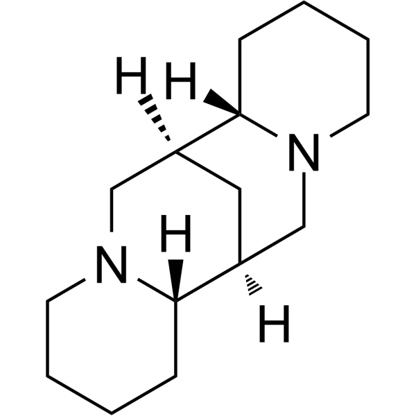 Sparteine
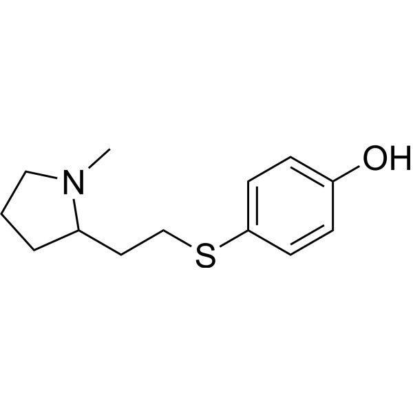 SIB-1553A free base
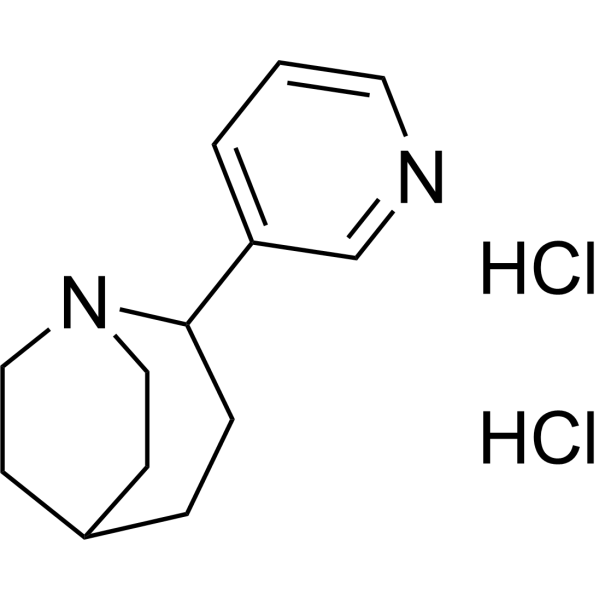 TC-1698 dihydrochloride
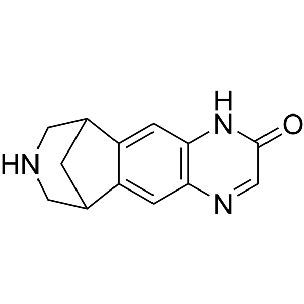 Hydroxy Varenicline
InvivoChem的所有产品仅用于作科学研究,不面向患者销售
Copyright 2020 InvivoChem LLC | All Rights Reserved 粤ICP备20063088号-1


