
| 规格 | 价格 | 库存 | 数量 |
|---|---|---|---|
| 5mg |
|
||
| 10mg |
|
||
| 25mg |
|
||
| 50mg |
|
||
| 100mg |
|
||
| 250mg |
|
||
| Other Sizes |
|
| 靶点 |
Chk1 (IC50 = 3 nM); Chk2 (IC50 = 1500 nM); CDK2 (IC50 = 160 nM)
|
|---|---|
| 体外研究 (In Vitro) |
体外活性:SCH 900776 是一种效力较低的 Chk2 和 CDK2 抑制剂,IC50 分别为 1.5 μM 和 0.16 μM。 SCH 900776 对细胞色素 P450 人肝微粒体亚型 1A2、2C9、2C19、2D6 和 3A4 无显着抑制作用。接触羟基脲 24 小时后,SCH 900776 会导致 DNA 复制能力出现剂量依赖性丧失。 SCH 900776 增强羟基脲、5-氟尿嘧啶和阿糖胞苷的 γ-H2AX 反应。与抗代谢药结合使用,SCH 900776 可在 2 小时内诱导 γ-H2AX 积累,表明复制叉崩溃和双链 DNA 断裂。此外,SCH 900776 以剂量依赖性方式抑制 Chk1 pS296 自磷酸化的积累。增殖的 WS1 细胞暴露于 SCH 900776 与 Chk1 pS345 的快速、剂量依赖性积累相关,表明正常细胞的循环群体在暴露于 SCH 900776 后诱导 Chk1 pS345 作为无效循环的一部分,可能是由 AT 家族激酶驱动的和DNA-PK。激酶测定:激酶分析器服务用于生成 SCH 900776 针对各种丝氨酸/苏氨酸和酪氨酸激酶的一般选择性数据。测定通常在两种浓度的 SCH 900776(0.5 和 5 μM)和固定浓度(10 μM)的 ATP 下进行。细胞测定:在体外,SCH 900776 以剂量依赖性方式阻断 Chk1 pS296 自磷酸化的积累。研究发现,用 SCH 900776 处理增殖的 WS1 细胞与 Chk1 pS345 的快速、剂量依赖性积累相关,这表明正常细胞的循环群体会响应 SCH 900776 的抑制而诱导 Chk1 pS345,作为无效周期 1 的一部分。
|
| 体内研究 (In Vivo) |
吉西他滨给药 30 分钟后,4 mg/kg SCH 900776 足以诱导 γ-H2AX 生物标志物,而相对于单独使用吉西他滨或 SCH 900776,8 mg/kg 则可增强肿瘤药效学和消退反应。 SCH 900776 的剂量递增(16 mg/kg 和 32 mg/kg)可导致肿瘤反应逐渐改善。重要的是,SCH 900776 的剂量与稳健的生物标志物激活和改善的肿瘤反应相关,但与吉西他滨对 BALB/c 小鼠血液学参数的毒性增强无关。
|
| 酶活实验 |
SCH 900776 针对各种丝氨酸/苏氨酸和酪氨酸激酶的一般选择性数据是通过激酶分析器服务生成的。 SCH 900776 通常用于两种浓度(0.5 和 5 μM)以及固定浓度 (10 μM) ATP 的测定。
以基于CDC25C的生物素化肽为底物,杆状病毒表达系统表达的重组His-Chk1为酶源的体外实验。在含有 50 mM Tris pH8.0、10 mM MgCl2 和 1 mM DTT 的激酶缓冲液中,His-Chk1 被稀释至 32 nM。将 CDC25C(CDC25 Ser216 C 末端生物素化肽)肽在激酶缓冲液中稀释至浓度 1.93 μM。为了在添加起始溶液后产生 6.2 nM Chk1、385 nM CDC25C 和 1% DMSO 的最终反应浓度,将 20 μL 32 nM Chk1 酶溶液和 20 μL 1.926 μM CDC25C 混合并与 10对于每个激酶反应,用 10% DMSO 稀释 μL SCH 900776。添加 50 μL 起始溶液(其中含有 2 μM ATP 和 0.2 μCi 33P-ATP)启动反应,最终反应浓度为 1 μM ATP 和 0.2 μCi 每个反应 33 P-ATP。激酶反应在室温下运行 2 小时,并通过添加 100 μL 终止液来终止,终止液由 2 M NaCl、1% H3PO4 和 5 组成。 mg/mL 链霉亲和素包被的 SPA 珠。 Filtermate 通用收集器与 96 孔 GF/B 过滤板组合用于收集 SPA 珠子。 2 M NaCl 和 2 M NaCl 加 1% 磷酸均用于洗珠两次。之后,用 TopCount 96 孔液体闪烁计数器测量信号。使用 SCH 900776 在八个点上连续稀释一式两份来创建剂量反应曲线。通过使用非线性回归分析,获得IC50值。 激酶测定[1] CHK1、CHK2和CDK激酶测定已在前面描述。Millipore激酶分析器服务用于生成SCH 900776对多种丝氨酸/苏氨酸和酪氨酸激酶的一般选择性数据。通常在两种浓度的SCH 900776(0.5和5μmol/L)和固定浓度的ATP(10μmol/L)下进行检测。数据以剩余活动百分比的形式提供,与未受抑制的对照组相比。 使用温度依赖性荧光进行亲和力评估[1] 在白色96孔PCR板中,将1μmol/L CHK1重组激酶结构域蛋白(氨基酸残基2-274)与微摩尔浓度(通常为1-50μmol/L)的化合物在20μL测定缓冲液(25 mmol/L HEPES,pH 7.4,300 mmol/L NaCl,5 mmol/L二硫苏糖醇,2%二甲亚砜,Sypro Orange 5x)中混合。该板用透明条密封,并放置在热循环仪中。在25°C至95°C的熔化过程中,每0.5°C的增量监测一次荧光强度。数据被导出到Excel中,并经过专有的自定义曲线拟合算法(未公布),以得出温度依赖性荧光(TdF)Kd值。对于CHK1 TdF数据,通常使用双态结合模型(化合物结合天然和热展开的熔融球态)。与靶激酶的熔融球态结合的化合物通常比天然状态弱1000倍以上。由于结合焓变化的不确定性,所有TdF Kd值的误差幅度约为50%。 |
| 细胞实验 |
为了进行细胞生长测定,将 500-1000 个细胞以低密度接种到 96 孔板中,然后用药物处理细胞一整天(每个浓度 8 个孔)。处理后,冲洗细胞并在37°C的新培养基中培养5至7天。在细胞达到汇合之前,将细胞裂解、清洗并用 Hoechst 33258 染色。使用微孔板分光荧光计测量荧光。用抑制50%生长的药物浓度的平均值和标准误来表示结果。
γ-H2AX测定[1] 简单地说,将细胞暴露于抗代谢物中以诱导CHK1的激活。对照人群不进行治疗。然后在2小时的暴露窗口(在抗代谢物存在的情况下)将 SCH 900776 滴定到细胞上。在SCH 900776共暴露2小时后,将细胞固定并渗透(70%乙醇),然后用异硫氰酸荧光素(FITC)偶联抗γ- h2ax单克隆抗体染色。细胞用碘化丙啶反染,随后使用流式细胞仪(Becton Dickinson LSR II)或Discovery 1免疫荧光平台进行分析。实验通常进行三次,数据以γ-H2AX阳性细胞的百分比表示,从而反映了γ-H2AX表型的总体外显率。 用活性caspase>评价凋亡诱导 [1] SCH 900776。然后清洗细胞,去除所有抗代谢物和SCH 900776。在这一点(T0或释放)评估Caspase活性,并在T + 24和T + 48小时进行进一步的检测。细胞随后用荧光标记的caspase底物孵育;细胞内底物的摄取和荧光与激活的半胱天冬酶水平相关。然后用流式细胞术测定表达活化半胱天冬酶的细胞百分比。 溴脱氧尿苷掺入试验[1] 将细胞镀于10厘米的组织培养皿中,使其粘附。将细胞暴露于不同浓度的SCH 900776中超过2小时,无论事先是否有抗代谢物暴露。然后清洗细胞,并允许24小时尝试恢复s期。随后短暂(30分钟)暴露于溴脱氧尿苷(BrdU),以评估能够以活的方式重新进入细胞周期的细胞百分比。然后收获细胞,固定并渗透。随后进行酸变性步骤,以暴露基因组DNA中合并的BrdU表位,之后用fitc偶联的BrdU特异性单克隆抗体对样品进行免疫染色。然后用碘化丙啶对细胞进行反染色,以评估DNA含量并使用流式细胞术进行分析。BrdU阳性染色和碘化丙啶信号的双变量分析可以评估正在进行DNA合成的细胞数量和细胞系的整体细胞周期分布(G1, S, G2-M和亚G1)。每个浓度下每个种群的百分比. |
| 动物实验 |
Female nude mice injected subcutaneously with A2780 or MiaPaCa2 cells
~50 mg/kg Administered intraperitoneally Certain cell lines are grown in vitro, once they have been washed with PBS, they are resuspended in 50% Matrigel in PBS to a final concentration of 4×10⁷ to 5×10⁷ cells per mL for tumor implantation. A subcutaneous injection of 0.1 mL of this suspension is given to naked mice in the flank area. Tumor length (L), width (W), and height (H) are measured twice a week on each mouse using a caliper. The tumor volume is then determined using the formula (L×W×H)/2. Ten animals are randomly assigned to treatment groups and given intraperitoneally either SCH 900776 (formulated in 20% hydroxypropyl β-cyclodextrin) or specific chemotherapeutic agents, prepared in accordance with recommended guidelines. Measurements are taken both during and after the treatment phases of the tumor volumes and body masses. Prior to normalization to starting volume, data are recorded as means±SEM. In some experiments, time to progression to 10x starting volume, or TTP 10x, is recorded. The tumor and surrounding skin are removed during necropsy, preserved for an overnight period in 10% formalin, and then cleaned and preserved in 70% ethanol for pharmacodynamic marker analyses in mice. Shave an area about 4 square inches in size in order to perform skin punch biopsies. In order to induce local anesthesia in dogs, lidocaine is administered subcutaneously, while inhaled isofluorane is used for anesthesia in rats. Using a 4 mm biopsy punch, samples are obtained. Before being cleaned or stored in 70% ethanol, skin punches are fixed in 10% formalin for an entire night. In vivo tumor growth assessments, sampling, and skin biopsies [1] For tumor implantation, specific cell lines were grown in vitro, washed once with PBS and resuspended in 50% Matrigel (BD Biosciences) in PBS to a final concentration of 4 × 10⁷ to 5 × 10⁷ cells per mL. Nude mice were injected with 0.1 mL of this suspension subcutaneously in the flank region. Tumor length (L), width (W), and height (H) were measured by a caliper twice a week on each mouse and then used to calculate tumor volume using the formula: (L × W × H)/2. Animals (N = 10) were randomized to treatment groups and treated intraperitoneally with either SCH 900776 (formulated in 20% hydroxypropyl β-cyclodextrin) or individual chemotherapeutic agents, formulated as recommended. Tumor volumes and body weights were measured during and after the treatment periods. Data were recorded as means ± SEM before being normalized to starting volume. Time to progression to 10x starting volume (TTP 10x) was monitored in some experiments. Animals were euthanized according to Institutional Animal Care and Use Committee guidelines. For pharmacodynamic marker analyses in mice, tumors and adjacent skin were collected at necropsy, fixed overnight in 10% formalin, and washed/stored in 70% ethanol. For skin punch biopsies, an area of approximately 4 square inches was shaved. Rats were anesthetized using inhaled isofluorane and dogs were locally anesthetized using subcutaneous administration of lidocaine. Samples were collected using a 4 mm biopsy punch. Skin punches were fixed in 10% formalin overnight before washing/storage in 70% ethanol. Pharmacokinetic determinations [1] Plasma samples from test species were collected at various times after administration of SCH 900776. At each time-point, blood samples from 3 animals were combined and analyzed for SCH 900776 by LC/MS. Pharmacokinetic variables were estimated from the plasma concentration data. Cmax values (maximum plasma concentration) were taken directly from the plasma concentration-time profiles, and the area under the plasma concentration versus time curve area under curve (AUC) was calculated using the linear trapezoidal rule. |
| 参考文献 | |
| 其他信息 |
Sch 900776 has been used in trials studying the treatment of Neoplasms, Hodgkin Disease, Adult Erythroleukemia, Lymphoma, Non-Hodgkin, and Myelogenous Leukemia, Acute, among others.
CHK1 Inhibitor MK-8776 is an agent targeting cell cycle checkpoint kinase 1 (Chk1) with potential radiosensitization and chemosensitization activities. Chk1 inhibitor MK-8776 specifically binds to and inhibits Chk1, which may result in tumor cells bypassing Chk1-dependent cell cycle arrest in the S and G2/M phases to undergo DNA repair prior to entry into mitosis; tumor cells may thus be sensitized to the DNA-damaging effects of ionizing radiation and alkylating chemotherapeutic agents. Chk1 is an ATP-dependent serine-threonine kinase that in response to DNA damage phosphorylates cdc25 phosphatases, resulting in inhibitory tyrosine phosphorylation of CDK-cyclin complexes and cell cycle arrest. 6-bromo-3-(1-methyl-4-pyrazolyl)-5-(3-piperidinyl)-7-pyrazolo[1,5-a]pyrimidinamine is a pyrazolopyrimidine. See also: Mk-8776 (annotation moved to). Checkpoint kinase 1 (CHK1) is an essential serine/threonine kinase that responds to DNA damage and stalled DNA replication. CHK1 is essential for maintenance of replication fork viability during exposure to DNA antimetabolites. In human tumor cell lines, ablation of CHK1 function during antimetabolite exposure led to accumulation of double-strand DNA breaks and cell death. Here, we extend these observations and confirm ablation of CHK2 does not contribute to these phenotypes and may diminish them. Furthermore, concomitant suppression of cyclin-dependent kinase (CDK) activity is sufficient to completely antagonize the desired CHK1 ablation phenotypes. These mechanism-based observations prompted the development of a high-content, cell-based screen for γ-H2AX induction, a surrogate marker for double-strand DNA breaks. This mechanism-based functional approach was used to optimize small molecule inhibitors of CHK1. Specifically, the assay was used to mechanistically define the optimal in-cell profile with compounds exhibiting varying degrees of CHK1, CHK2, and CDK selectivity. Using this approach, SCH 900776 was identified as a highly potent and functionally optimal CHK1 inhibitor with minimal intrinsic antagonistic properties. SCH 900776 exposure phenocopies short interfering RNA-mediated CHK1 ablation and interacts synergistically with DNA antimetabolite agents in vitro and in vivo to selectively induce dsDNA breaks and cell death in tumor cell backgrounds. [1] Many anticancer agents damage DNA and arrest cell-cycle progression primarily in S or G(2) phase of the cell cycle. Previous studies with the topoisomerase I inhibitor SN38 have shown the efficacy of the Chk1 inhibitor UCN-01 to overcome this arrest and induce mitotic catastrophe. UCN-01 was limited in clinical trials by unfavorable pharmacokinetics. SCH900776 is a novel and more selective Chk1 inhibitor that potently inhibits Chk1 and abrogates cell-cycle arrest induced by SN38. Like UCN-01, abrogation of SN38-induced arrest enhances the rate of cell death but does not increase overall cell death. In contrast, SCH900776 reduced the growth-inhibitory concentration of hydroxyurea by 20- to 70-fold. A similar magnitude of sensitization was observed with cytarabine. A 5- to 10-fold sensitization occurred with gemcitabine, but no sensitization occurred with cisplatin, 5-fluorouracil, or 6-thioguanine. Sensitization occurred at hydroxyurea concentrations that marginally slowed DNA replication without apparent activation of Chk1, but this led to dependence on Chk1 that increased with time. For example, when added 18 hours after hydroxyurea, SCH900776 induced DNA double-strand breaks consistent with rapid collapse of replication forks. In addition, some cell lines were highly sensitive to SCH900776 alone, and these cells required lower concentrations of SCH900776 to sensitize them to hydroxyurea. We conclude that some tumors may be very sensitive to the combination of SCH900776 and hydroxyurea. Delayed administration of SCH900776 may be more effective than concurrent treatment. SCH900776 is currently in phase I clinical trials, and these results provide the rationale and schedule for future clinical trials.[2] Purpose: Previous studies have shown that the replication checkpoint, which involves the kinases ataxia telangiectasia mutated and Rad3 related (ATR) and Chk1, contributes to cytarabine resistance in cell lines. In the present study, we examined whether this checkpoint is activated in clinical acute myelogenous leukemia (AML) during cytarabine infusion in vivo and then assessed the impact of combining cytarabine with the recently described Chk1 inhibitor SCH 900776 in vitro. Experimental design: AML marrow aspirates harvested before and during cytarabine infusion were examined by immunoblotting. Human AML lines treated with cytarabine in the absence or presence of SCH 900776 were assayed for checkpoint activation by immunoblotting, nucleotide incorporation into DNA, and flow cytometry. Long-term effects in AML lines, clinical AML isolates, and normal myeloid progenitors were assayed using clonogenic assays. Results: Immunoblotting revealed increased Chk1 phosphorylation, a marker of checkpoint activation, in more than half of Chk1-containing AMLs after 48 hours of cytarabine infusion. In human AML lines, SCH 900776 not only disrupted cytarabine-induced Chk1 activation and S-phase arrest but also markedly increased cytarabine-induced apoptosis. Clonogenic assays demonstrated that SCH 900776 enhanced the antiproliferative effects of cytarabine in AML cell lines and clinical AML samples at concentrations that had negligible impact on normal myeloid progenitors. Conclusions: These results not only provide evidence for cytarabine-induced S-phase checkpoint activation in AML in the clinical setting, but also show that a selective Chk1 inhibitor can overcome the S-phase checkpoint and enhance the cytotoxicity of cytarabine. Accordingly, further investigation of the cytarabine/SCH 900776 combination in AML appears warranted.[3] |
| 分子式 |
C15H18BRN7
|
|
|---|---|---|
| 分子量 |
376.25
|
|
| 精确质量 |
375.08
|
|
| 元素分析 |
C, 47.88; H, 4.82; Br, 21.24; N, 26.06
|
|
| CAS号 |
891494-63-6
|
|
| 相关CAS号 |
SCH900776 (S-isomer);891494-64-7
|
|
| PubChem CID |
46239015
|
|
| 外观&性状 |
white to off-white Solid powder
|
|
| 密度 |
1.8±0.1 g/cm3
|
|
| 折射率 |
1.819
|
|
| LogP |
0.76
|
|
| tPSA |
86.06
|
|
| 氢键供体(HBD)数目 |
2
|
|
| 氢键受体(HBA)数目 |
5
|
|
| 可旋转键数目(RBC) |
2
|
|
| 重原子数目 |
23
|
|
| 分子复杂度/Complexity |
425
|
|
| 定义原子立体中心数目 |
1
|
|
| SMILES |
NC1=C(Br)C([C@H]2CNCCC2)=NC2=C(C3=CN(C)N=C3)C=NN12
|
|
| InChi Key |
GMIZZEXBPRLVIV-SECBINFHSA-N
|
|
| InChi Code |
InChI=1S/C15H18BrN7/c1-22-8-10(6-19-22)11-7-20-23-14(17)12(16)13(21-15(11)23)9-3-2-4-18-5-9/h6-9,18H,2-5,17H2,1H3/t9-/m1/s1
|
|
| 化学名 |
6-bromo-3-(1-methylpyrazol-4-yl)-5-[(3R)-piperidin-3-yl]pyrazolo[1,5-a]pyrimidin-7-amine
|
|
| 别名 |
|
|
| HS Tariff Code |
2934.99.9001
|
|
| 存储方式 |
Powder -20°C 3 years 4°C 2 years In solvent -80°C 6 months -20°C 1 month |
|
| 运输条件 |
Room temperature (This product is stable at ambient temperature for a few days during ordinary shipping and time spent in Customs)
|
| 溶解度 (体外实验) |
|
|||
|---|---|---|---|---|
| 溶解度 (体内实验) |
配方 1 中的溶解度: ≥ 2.5 mg/mL (6.64 mM) (饱和度未知) in 10% DMSO + 40% PEG300 + 5% Tween80 + 45% Saline (这些助溶剂从左到右依次添加,逐一添加), 澄清溶液。
例如,若需制备1 mL的工作液,可将100 μL 25.0 mg/mL澄清DMSO储备液加入到400 μL PEG300中,混匀;然后向上述溶液中加入50 μL Tween-80,混匀;加入450 μL生理盐水定容至1 mL。 *生理盐水的制备:将 0.9 g 氯化钠溶解在 100 mL ddH₂O中,得到澄清溶液。 配方 2 中的溶解度: ≥ 2.5 mg/mL (6.64 mM) (饱和度未知) in 10% DMSO + 90% (20% SBE-β-CD in Saline) (这些助溶剂从左到右依次添加,逐一添加), 澄清溶液。 例如,若需制备1 mL的工作液,可将 100 μL 25.0 mg/mL澄清DMSO储备液加入900 μL 20% SBE-β-CD生理盐水溶液中,混匀。 *20% SBE-β-CD 生理盐水溶液的制备(4°C,1 周):将 2 g SBE-β-CD 溶解于 10 mL 生理盐水中,得到澄清溶液。 View More
配方 3 中的溶解度: ≥ 2.5 mg/mL (6.64 mM) (饱和度未知) in 10% DMSO + 90% Corn Oil (这些助溶剂从左到右依次添加,逐一添加), 澄清溶液。 配方 4 中的溶解度: 4% DMSO +30% Propylene glycol : 5 mg/mL 1、请先配制澄清的储备液(如:用DMSO配置50 或 100 mg/mL母液(储备液)); 2、取适量母液,按从左到右的顺序依次添加助溶剂,澄清后再加入下一助溶剂。以 下列配方为例说明 (注意此配方只用于说明,并不一定代表此产品 的实际溶解配方): 10% DMSO → 40% PEG300 → 5% Tween-80 → 45% ddH2O (或 saline); 假设最终工作液的体积为 1 mL, 浓度为5 mg/mL: 取 100 μL 50 mg/mL 的澄清 DMSO 储备液加到 400 μL PEG300 中,混合均匀/澄清;向上述体系中加入50 μL Tween-80,混合均匀/澄清;然后继续加入450 μL ddH2O (或 saline)定容至 1 mL; 3、溶剂前显示的百分比是指该溶剂在最终溶液/工作液中的体积所占比例; 4、 如产品在配制过程中出现沉淀/析出,可通过加热(≤50℃)或超声的方式助溶; 5、为保证最佳实验结果,工作液请现配现用! 6、如不确定怎么将母液配置成体内动物实验的工作液,请查看说明书或联系我们; 7、 以上所有助溶剂都可在 Invivochem.cn网站购买。 |
| 制备储备液 | 1 mg | 5 mg | 10 mg | |
| 1 mM | 2.6578 mL | 13.2890 mL | 26.5781 mL | |
| 5 mM | 0.5316 mL | 2.6578 mL | 5.3156 mL | |
| 10 mM | 0.2658 mL | 1.3289 mL | 2.6578 mL |
1、根据实验需要选择合适的溶剂配制储备液 (母液):对于大多数产品,InvivoChem推荐用DMSO配置母液 (比如:5、10、20mM或者10、20、50 mg/mL浓度),个别水溶性高的产品可直接溶于水。产品在DMSO 、水或其他溶剂中的具体溶解度详见上”溶解度 (体外)”部分;
2、如果您找不到您想要的溶解度信息,或者很难将产品溶解在溶液中,请联系我们;
3、建议使用下列计算器进行相关计算(摩尔浓度计算器、稀释计算器、分子量计算器、重组计算器等);
4、母液配好之后,将其分装到常规用量,并储存在-20°C或-80°C,尽量减少反复冻融循环。
计算结果:
工作液浓度: mg/mL;
DMSO母液配制方法: mg 药物溶于 μL DMSO溶液(母液浓度 mg/mL)。如该浓度超过该批次药物DMSO溶解度,请首先与我们联系。
体内配方配制方法:取 μL DMSO母液,加入 μL PEG300,混匀澄清后加入μL Tween 80,混匀澄清后加入 μL ddH2O,混匀澄清。
(1) 请确保溶液澄清之后,再加入下一种溶剂 (助溶剂) 。可利用涡旋、超声或水浴加热等方法助溶;
(2) 一定要按顺序加入溶剂 (助溶剂) 。
| NCT Number | Recruitment | interventions | Conditions | Sponsor/Collaborators | Start Date | Phases |
| NCT00779584 | Completed | Drug: MK-8776 Drug: Gemcitabine |
Hodgkin Disease Neoplasms |
Merck Sharp & Dohme LLC | October 17, 2008 | Phase 1 |
| NCT01870596 | Completed | Drug: CHK1 Inhibitor SCH 900776 Drug: Cytarabine |
Adult Acute Monocytic Leukemia Adult Erythroleukemia |
National Cancer Institute (NCI) |
May 2013 | Phase 2 |
| NCT00907517 | Completed | Drug: MK-8776 Drug: Cytarabine |
Myelogenous Leukemia, Acute Leukemia, Lymphocytic, Acute |
Merck Sharp & Dohme LLC | July 29, 2009 | Phase 1 |
") Comparative efficacy of UCN-01 and SCH900776 at inhibiting Chk1 and abrogating SN38-induced cell cycle arrest.Mol Cancer Ther.2012 Feb;11(2):427-38. |
|---|
") The impact of checkpoint inhibitors on sensitivity of cells to SN38, hydroxyurea and cisplatin.Mol Cancer Ther.2012 Feb;11(2):427-38. |
") Analysis of cell cycle perturbation induced by hydroxyurea.Mol Cancer Ther.2012 Feb;11(2):427-38. |
") Impact of concentration and schedule of hydroxyurea and SCH900776 on DNA damage and cytotoxicityA.Mol Cancer Ther.2012 Feb;11(2):427-38. |
|---|
") Comparative efficacy of UCN-01 and SCH900776 at abrogating SN38-induced cell cycle arrest in cells suppressed for Chk1.A.MDA-MB-231ΔChk1 cells were incubated with 10 ng/mL SN38 for 24 h.Mol Cancer Ther.2012 Feb;11(2):427-38. |
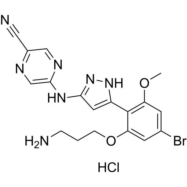 CHK1-IN-4 hydrochloride
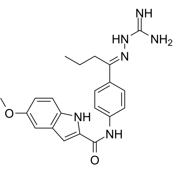 PV1162
 CHK1-IN-9
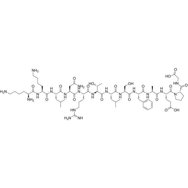 Ziptide
InvivoChem的所有产品仅用于作科学研究,不面向患者销售
Copyright 2020 InvivoChem LLC | All Rights Reserved 粤ICP备20063088号-1


 COA
COA
