
| 规格 | 价格 | 库存 | 数量 |
|---|---|---|---|
| 1mg |
|
||
| 5mg |
|
||
| 10mg |
|
||
| 25mg |
|
||
| 50mg |
|
||
| 100mg |
|
||
| 250mg |
|
||
| Other Sizes |
|
| 靶点 |
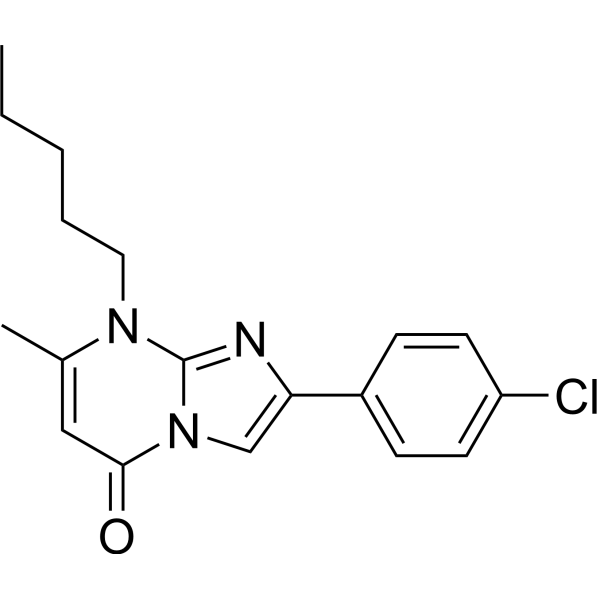
ACVR1 (IC50 = 5 nM); BMPR1A (IC50 = 30 nM); ALK2 (IC50 = 5 nM), ALK3 (IC50 = 30 nM)
|
||
|---|---|---|---|
| 体外研究 (In Vitro) |
LDN-193189 的 IC50 值分别为 5 nM 和 30 nM,可有效抑制 BMP I 型受体 ALK2 和 ALK3 的转录活性[1]。 LDN-193189 的 IC50 值低于 500 nM,对激活素和 TGF-β I 型受体 ALK4、ALK5 和 ALK7 的影响可以忽略不计[1]。 ActRIIA 与 LDN-193189 结合,Kd 值为 14 nM[2]。 LDN-193189(0.5 μM;30 分钟)靶向抑制 GDF8 触发的肌源性转录因子和 Smad2/3 信号传导[2]。有效抑制 GDF8 诱导的 Smad3/4 报告基因活性的是 LDN-193189(0.05、0.5 和 5 μM)[2]。 GDF8 处理的成肌细胞看到 LDN-193189 (0–5 μM) 恢复了其肌生成[2]。
|
||
| 体内研究 (In Vivo) |
LDN-193189(腹腔注射;3 mg/kg;每天;35 天)可能会影响乳腺癌细胞与其周围骨骼的相互作用[3]。 LDN-193189(腹腔注射;3 mg/kg;单剂量)可减少功能损伤和异位骨化 [1]。
BMP I型受体激酶的选择性抑制剂LDN-193189(参考文献6)抑制由腺病毒特异性Cre诱导的表达caALK2的组织中BMP信号效应器SMAD1、SMAD5和SMAD8的激活(Ad.Cre)。这种治疗减少了异位骨化和功能损害。与Ad.Cre对caALK2的局部诱导(这会引起炎症)相反,caALK2在出生后的整体表达(在不使用Ad.Cre的情况下诱导,因此没有炎症)不会导致异位骨化。然而,如果在这种情况下,用对照腺病毒提供炎症刺激,则会诱导异位骨形成。与LDN-193189一样,皮质类固醇抑制Ad.Cre-injected突变小鼠的骨化,表明caALK2表达和炎症环境都是该模型异位骨化发展所必需的。这些结果支持ALK2激酶活性失调在FOP发病机制中的作用,并表明小分子抑制BMP I型受体活性可能有助于治疗FOP和与过量BMP信号相关的异位骨化综合征。[1] 在本研究中,研究人员旨在调查LDN-193189化合物(BMP I型受体的强效抑制剂)对体内转移发展的影响。将ZNF217-revLuc细胞注射到裸鼠(n=16)的左心室中,而对照组小鼠(n=13)接种对照pcDNA6-revLuc电池。每组小鼠用LDN-193189治疗或不治疗35天。我们发现,对小鼠进行全身LDN-193189治疗,通过增加转移的数量和大小,显著促进了转移的发展。在注射pcDNA6-revLuc的小鼠中,LDN-193189也影响了转移出现的动力学。总之,这些数据表明,在体内,LDN-193189可能影响癌症细胞与骨环境之间的相互作用,有利于多发转移的出现和发展。因此,我们的报告强调了骨转移治疗中药物选择和治疗策略的重要性。[3] |
||
| 酶活实验 |
Id1和纤溶酶原激活物抑制剂-1启动子萤光素酶报告基因测定[1]
我们使用Fugene6,用0.3μg Id1启动子荧光素酶报告子构建体(BRE-Luc30,由P.ten Dijke提供)和0.6μg表达组成型活性形式的BMP I型受体(caALK2、caALK3或caALK631,由K.Miyazono提供)的质粒在六孔板中瞬时转染生长至50%汇合的小鼠PASMC。为了评估激活素和TGF-βI型受体的功能,我们用0.3μg PAI1(纤溶酶原激活物抑制剂-1)启动子萤光素酶报告子构建体(CAGA-Luc32,由P.ten Dijke提供)与0.6μg表达I型受体组成型活性形式(caALK4、caALK5和caALK733)的质粒联合瞬时转染PASMC。对于两个报告质粒,我们使用0.2μg的pRL-TK雷尼拉萤光素酶来控制转染效率。我们在转染后1小时开始用LDN-193189(2 nM–32μM)或载体孵育PASMC。我们采集了细胞提取物,并用双荧光素酶测定试剂盒通过萤火虫与雷尼拉萤光素酶活性的比率来量化相对启动子活性。
|
||
| 细胞实验 |
Smad1、Smad5和Smad8磷酸化的免疫印迹分析[1]
我们在SDS裂解缓冲液(62.5mM Tris-HCl(pH 6.8)、2%SDS、10%甘油、50mM二硫苏糖醇和0.01%溴酚蓝)中机械匀浆细胞提取物,通过SDS-PAGE分离蛋白质,用磷酸化Smad1、Smad5和Smad8特异性多克隆抗体、磷酸化Smad2或兔Smad1或Smad2特异性单克隆抗体免疫印迹,并用ECL-Plus观察免疫反应蛋白。
|
||
| 动物实验 |
|
||
| 参考文献 |
|
||
| 其他信息 |
Fibrodysplasia ossificans progressiva (FOP) is a congenital disorder of progressive and widespread postnatal ossification of soft tissues and is without known effective treatments. Affected individuals harbor conserved mutations in the ACVR1 gene that are thought to cause constitutive activation of the bone morphogenetic protein (BMP) type I receptor, activin receptor-like kinase-2 (ALK2). Here we show that intramuscular expression in the mouse of an inducible transgene encoding constitutively active ALK2 (caALK2), resulting from a glutamine to aspartic acid change at amino acid position 207, leads to ectopic endochondral bone formation, joint fusion and functional impairment, thus phenocopying key aspects of human FOP. A selective inhibitor of BMP type I receptor kinases, LDN-193189 (ref. 6), inhibits activation of the BMP signaling effectors SMAD1, SMAD5 and SMAD8 in tissues expressing caALK2 induced by adenovirus specifying Cre (Ad.Cre). This treatment resulted in a reduction in ectopic ossification and functional impairment. In contrast to localized induction of caALK2 by Ad.Cre (which entails inflammation), global postnatal expression of caALK2 (induced without the use of Ad.Cre and thus without inflammation) does not lead to ectopic ossification. However, if in this context an inflammatory stimulus was provided with a control adenovirus, ectopic bone formation was induced. Like LDN-193189, corticosteroid inhibits ossification in Ad.Cre-injected mutant mice, suggesting caALK2 expression and an inflammatory milieu are both required for the development of ectopic ossification in this model. These results support the role of dysregulated ALK2 kinase activity in the pathogenesis of FOP and suggest that small molecule inhibition of BMP type I receptor activity may be useful in treating FOP and heterotopic ossification syndromes associated with excessive BMP signaling.[1]
GDF8, or myostatin, is a member of the TGF-β superfamily of secreted polypeptide growth factors. GDF8 is a potent negative regulator of myogenesis both in vivo and in vitro. We found that GDF8 signaling was inhibited by the small molecule ATP competitive inhibitors dorsomorphin and LDN-193189. These compounds were previously shown to be potent inhibitors of BMP signaling by binding to the BMP type I receptors ALK1/2/3/6. We present the crystal structure of the type II receptor ActRIIA with dorsomorphin and demonstrate that dorsomorphin or LDN-193189 target GDF8 induced Smad2/3 signaling and repression of myogenic transcription factors. As a result, both inhibitors rescued myogenesis in myoblasts treated with GDF8. As revealed by quantitative live cell microscopy, treatment with dorsomorphin or LDN-193189 promoted the contractile activity of myotubular networks in vitro. We therefore suggest these inhibitors as suitable tools to promote functional myogenesis.[2] Breast cancer with bone metastasis is essentially incurable with current anticancer therapies. The bone morphogenetic protein (BMP) pathway is an attractive therapeutic candidate, as it is involved in the bone turnover and in cancer cell formation and their colonization of distant organs such as the bone. We previously reported that in breast cancer cells, the ZNF217 oncogene drives BMP pathway activation, increases the metastatic growth rate in the bone, and accelerates the development of severe osteolytic lesions in mice. [3] |
| 分子式 |
C25H24CL2N6
|
|
|---|---|---|
| 分子量 |
479.4
|
|
| 精确质量 |
478.143
|
|
| 元素分析 |
C, 62.63; H, 5.05; Cl, 14.79; N, 17.53
|
|
| CAS号 |
1435934-00-1
|
|
| 相关CAS号 |
LDN193189;1062368-24-4;LDN193189 Tetrahydrochloride;2310134-98-4
|
|
| PubChem CID |
91900717
|
|
| 外观&性状 |
Typically exists as orange to red solids at room temperature
|
|
| tPSA |
58.4Ų
|
|
| 氢键供体(HBD)数目 |
3
|
|
| 氢键受体(HBA)数目 |
5
|
|
| 可旋转键数目(RBC) |
3
|
|
| 重原子数目 |
33
|
|
| 分子复杂度/Complexity |
587
|
|
| 定义原子立体中心数目 |
0
|
|
| SMILES |
N1=C2C(C=CC=C2)=C(C2=C3N(N=C2)C=C(C2=CC=C(N4CCNCC4)C=C2)C=N3)C=C1.[H]Cl.[H]Cl
|
|
| InChi Key |
CMQXLLAILGGLRV-UHFFFAOYSA-N
|
|
| InChi Code |
InChI=1S/C25H22N6.2ClH/c1-2-4-24-22(3-1)21(9-10-27-24)23-16-29-31-17-19(15-28-25(23)31)18-5-7-20(8-6-18)30-13-11-26-12-14-30;;/h1-10,15-17,26H,11-14H2;2*1H
|
|
| 化学名 |
4-[6-(4-piperazin-1-ylphenyl)pyrazolo[1,5-a]pyrimidin-3-yl]quinoline;dihydrochloride
|
|
| 别名 |
|
|
| HS Tariff Code |
2934.99.9001
|
|
| 存储方式 |
Powder -20°C 3 years 4°C 2 years In solvent -80°C 6 months -20°C 1 month 注意: 请将本产品存放在密封且受保护的环境中,避免吸湿/受潮。 |
|
| 运输条件 |
Room temperature (This product is stable at ambient temperature for a few days during ordinary shipping and time spent in Customs)
|
| 溶解度 (体外实验) |
|
|||
|---|---|---|---|---|
| 溶解度 (体内实验) |
注意: 如下所列的是一些常用的体内动物实验溶解配方,主要用于溶解难溶或不溶于水的产品(水溶度<1 mg/mL)。 建议您先取少量样品进行尝试,如该配方可行,再根据实验需求增加样品量。
注射用配方
注射用配方1: DMSO : Tween 80: Saline = 10 : 5 : 85 (如: 100 μL DMSO → 50 μL Tween 80 → 850 μL Saline)(IP/IV/IM/SC等) *生理盐水/Saline的制备:将0.9g氯化钠/NaCl溶解在100 mL ddH ₂ O中,得到澄清溶液。 注射用配方 2: DMSO : PEG300 :Tween 80 : Saline = 10 : 40 : 5 : 45 (如: 100 μL DMSO → 400 μL PEG300 → 50 μL Tween 80 → 450 μL Saline) 注射用配方 3: DMSO : Corn oil = 10 : 90 (如: 100 μL DMSO → 900 μL Corn oil) 示例: 以注射用配方 3 (DMSO : Corn oil = 10 : 90) 为例说明, 如果要配制 1 mL 2.5 mg/mL的工作液, 您可以取 100 μL 25 mg/mL 澄清的 DMSO 储备液,加到 900 μL Corn oil/玉米油中, 混合均匀。 View More
注射用配方 4: DMSO : 20% SBE-β-CD in Saline = 10 : 90 [如:100 μL DMSO → 900 μL (20% SBE-β-CD in Saline)] 口服配方
口服配方 1: 悬浮于0.5% CMC Na (羧甲基纤维素钠) 口服配方 2: 悬浮于0.5% Carboxymethyl cellulose (羧甲基纤维素) 示例: 以口服配方 1 (悬浮于 0.5% CMC Na)为例说明, 如果要配制 100 mL 2.5 mg/mL 的工作液, 您可以先取0.5g CMC Na并将其溶解于100mL ddH2O中,得到0.5%CMC-Na澄清溶液;然后将250 mg待测化合物加到100 mL前述 0.5%CMC Na溶液中,得到悬浮液。 View More
口服配方 3: 溶解于 PEG400 (聚乙二醇400) 请根据您的实验动物和给药方式选择适当的溶解配方/方案: 1、请先配制澄清的储备液(如:用DMSO配置50 或 100 mg/mL母液(储备液)); 2、取适量母液,按从左到右的顺序依次添加助溶剂,澄清后再加入下一助溶剂。以 下列配方为例说明 (注意此配方只用于说明,并不一定代表此产品 的实际溶解配方): 10% DMSO → 40% PEG300 → 5% Tween-80 → 45% ddH2O (或 saline); 假设最终工作液的体积为 1 mL, 浓度为5 mg/mL: 取 100 μL 50 mg/mL 的澄清 DMSO 储备液加到 400 μL PEG300 中,混合均匀/澄清;向上述体系中加入50 μL Tween-80,混合均匀/澄清;然后继续加入450 μL ddH2O (或 saline)定容至 1 mL; 3、溶剂前显示的百分比是指该溶剂在最终溶液/工作液中的体积所占比例; 4、 如产品在配制过程中出现沉淀/析出,可通过加热(≤50℃)或超声的方式助溶; 5、为保证最佳实验结果,工作液请现配现用! 6、如不确定怎么将母液配置成体内动物实验的工作液,请查看说明书或联系我们; 7、 以上所有助溶剂都可在 Invivochem.cn网站购买。 |
| 制备储备液 | 1 mg | 5 mg | 10 mg | |
| 1 mM | 2.0859 mL | 10.4297 mL | 20.8594 mL | |
| 5 mM | 0.4172 mL | 2.0859 mL | 4.1719 mL | |
| 10 mM | 0.2086 mL | 1.0430 mL | 2.0859 mL |
1、根据实验需要选择合适的溶剂配制储备液 (母液):对于大多数产品,InvivoChem推荐用DMSO配置母液 (比如:5、10、20mM或者10、20、50 mg/mL浓度),个别水溶性高的产品可直接溶于水。产品在DMSO 、水或其他溶剂中的具体溶解度详见上”溶解度 (体外)”部分;
2、如果您找不到您想要的溶解度信息,或者很难将产品溶解在溶液中,请联系我们;
3、建议使用下列计算器进行相关计算(摩尔浓度计算器、稀释计算器、分子量计算器、重组计算器等);
4、母液配好之后,将其分装到常规用量,并储存在-20°C或-80°C,尽量减少反复冻融循环。
计算结果:
工作液浓度: mg/mL;
DMSO母液配制方法: mg 药物溶于 μL DMSO溶液(母液浓度 mg/mL)。如该浓度超过该批次药物DMSO溶解度,请首先与我们联系。
体内配方配制方法:取 μL DMSO母液,加入 μL PEG300,混匀澄清后加入μL Tween 80,混匀澄清后加入 μL ddH2O,混匀澄清。
(1) 请确保溶液澄清之后,再加入下一种溶剂 (助溶剂) 。可利用涡旋、超声或水浴加热等方法助溶;
(2) 一定要按顺序加入溶剂 (助溶剂) 。
Inhibitor binding to ActRII.J Biol Chem.2015 Feb 6;290(6):3390-404.
Dorsomorphin and LDN-193189 inhibit GDF8-induced signaling pathways in undifferentiated and in differentiated primary human myoblasts and in C2C12 premyoblasts.J Biol Chem.2015 Feb 6;290(6):3390-404. |
|---|
Ligand-specific effects of kinase inhibitors on Smad2/3 and Smad1/5 phosphorylation.
Dorsomorphin treatment facilitates myotube formation.J Biol Chem.2015 Feb 6;290(6):3390-404. |
Dorsomorphin and LDN-193189 efficiently inhibit GDF8 induced Smad3/4 reporter gene activity.J Biol Chem.2015 Feb 6;290(6):3390-404. |
Dorsomorphin and LDN-193189 counteract GDF8-induced repression of myogenic differentiation.J Biol Chem.2015 Feb 6;290(6):3390-404. |
|---|
Dorsomorphin and LDN-193189 promote the formation of a contractile myotube network.J Biol Chem.2015 Feb 6;290(6):3390-404. |
 P17 Peptide
 Stevia Powder
 DCN1-IN-2
 CBT-295
InvivoChem的所有产品仅用于作科学研究,不面向患者销售
Copyright 2020 InvivoChem LLC | All Rights Reserved 粤ICP备20063088号-1


 COA
COA


