
| 规格 | 价格 | 库存 | 数量 |
|---|---|---|---|
| 1mg |
|
||
| 5mg |
|
||
| 10mg |
|
||
| 25mg |
|
||
| 50mg |
|
||
| 100mg |
|
||
| 1g |
|
||
| Other Sizes |
|
| 靶点 |
Camptothecins; TOP I; topoisomerase I
|
|---|---|
| 体外研究 (In Vitro) |
抗体-药物偶联物可选择性、高效地将抗癌药物递送至肿瘤组织,具有显着的抗肿瘤功效和广泛的治疗窗[2]。使用具有不同生物学特性的五种 CRC 细胞系研究了 [fam-] trastuzumab deruxtecan 对 CRC 的抗肿瘤活性。首先检查了这些不同细胞系中 HER2 在 mRNA 和蛋白质水平上的表达。免疫印迹分析以及 RT 和实时聚合酶链反应 (PCR) 分析显示,所有 CRC 细胞系中 HER2 蛋白和 HER2 mRNA 的量均远小于 NCI-N87 细胞。 [fam-] trastuzumab deruxtecan 减弱了 NCI-N87 细胞的活力,与之前的结果一致,而所有五种 CRC 细胞系均表现出对该药物的耐药性。这些发现表明 HER2 蛋白的表达水平可能决定对 [fam-] 曲妥珠单抗 deruxtecan 的敏感性。
Dxd(ADC的Exatecan衍生物)是一种强效的DNA拓扑异构酶I抑制剂,用作与HER2靶向ADC的偶联药物(DS-8201a),IC50为0.31μM。IC50范围为1.43 nM至4.07 nM,Dxd对KPL-4、NCI-N87、SK-BR-3和MDA-MB-468的人癌症细胞系具有细胞毒性;然而,以Dxd为有效载荷的对照IgG-ADC对四种细胞系(表达HER2)没有抑制作用。IC50值分别为26.8、25.4和6.7 ng/mL,DS-8201a(有效载荷为Dxd)对HER2阳性KPL-4、NCI-N87和SK-BR-3细胞系表现出显著的抑制作用,但对MDA-MB-468没有这种抑制作用(IC50,>10000 ng/mL)[3]。 DS-8201a的结构如图1A所示DS-8201a是一种靶向HER2的ADC,由抗HER2抗体和拓扑异构酶I抑制剂DX-8951(Dxd)的衍生物组成,它们通过马来酰亚胺-甘油-苯丙氨酸-甘油(GGFG)肽接头结合在一起。在用还原剂三(2-羧乙基)膦盐酸盐(TCEP HCl)还原链间二硫键后,接头有效载荷通过半胱氨酸残基与抗体偶联。由于四肽被溶酶体酶(如组织蛋白酶B和L)分解,这些酶在肿瘤细胞中高度表达,因此假设DS-8201a被溶酶体酶切割并释放Dxd,Dxd在与HER2受体结合后特异性攻击肿瘤细胞中的靶分子,并在肿瘤细胞内内化。通过使用RPC,DS-8201a的DAR被确定为约8,这是传统链间半胱氨酸偶联的理论最大载药量。因此,在HIC图中观察到均匀的药物分布(图1B)。我们证实,Dxd在拓扑异构酶I的抑制活性方面比SN-38和DX-8951f更有效,这是通过拓扑异酶I介导的DNA松弛试验测量的(图1C)[3]。 DS-8201a[3] 对癌症细胞生长的抑制作用 将DS-8201a对癌症细胞生长的抑制活性与体外与Dxd结合的抗HER2 Ab和对照IgG–ADC对各种人类癌症细胞系的抑制活性进行比较。首先通过流式细胞术分析评估细胞系KPL-4、NCI-N87、SK-BR-3和MDA-MB-468细胞表面HER2的表达(图2A)。KPL-4、NCI-N87和SK-BR-3的相对MFI分别为95.7、101.6和56.2,表明HER2在细胞表面上明确表达,而MDA-MB-468的相对MFI为1.0,表明MDA-MB-465中没有表达。观察到DS-8201a对HER2阳性KPL-4、NCI-N87和SK-BR-3具有显著的细胞生长抑制活性,IC50值分别为26.8、25.4和6.7 ng/mL,而对MDA-MB-468没有这种抑制作用,IC50值>10000 ng/mL(图2B)。尽管抗HER2抗体显示出对NCI-N87和SK-BR-3的细胞生长抑制活性,但这些活性比DS-8201a弱得多;NCI-N87和SK-BR-3的IC50值分别为204.2和65.9 ng/mL(图2B)。此外,对照IgG ADC在四种细胞系中均未显示出细胞生长抑制活性(图2B),尽管所有四种细胞株对有效载荷Dxd都很敏感(IC50:1.43 nmol/L-4.07 nmol/L)。这些结果表明,药物与抗HER2抗体结合显著增强了DS-8201a的细胞生长抑制活性,并且DS-8201a对HER2阳性细胞系显示出靶向特异性生长抑制作用。 |
| 体内研究 (In Vivo) |
测试了 [fam-] 曲妥珠单抗 deruxtecan 在表达 HER2 的异种移植肿瘤模型中的疗效。首次通过免疫组化(IHC)证实HCT116-Mock、HCT116-H2L或HCT116-H2H细胞裸鼠皮下肿瘤中HER2蛋白表达水平。以3.0 mg/kg剂量施用[fam-]曲妥珠单抗deruxtecan显着抑制由HCT116-H2L或HCT116-H2H细胞形成的肿瘤的生长,但不抑制由HCT116-Mock细胞形成的肿瘤的生长。第 24 天时,与 PBS 载体相比,[fam-] trastuzumab deruxtecan 对 HCT116-H2L 和 HCT116-H2H 细胞的抑制程度分别为 60% 和 93%。[fam-] trastuzumab deruxtecan 治疗对身体没有影响三组小鼠中任意一组的体重。因此,这些发现表明,异种移植模型中肿瘤对[fam-]曲妥珠单抗deruxtecan的敏感性取决于HER2表达水平,并且这种治疗与明显的毒性无关。
DS-8201a(DXdis为有效载荷,10mg/kg,静脉注射)在HER2 IHC 1+/FISH阴性表达的HER2低表达ST565和ST313模型以及KPL4、JIMT-1和Capan-1的HER2阳性模型中显示出强大的抗肿瘤活性[3]。 体内抗肿瘤活性[3] 在HER2阳性NCI-N87异种移植物模型中评估了DS-8201a的体内抗肿瘤活性DS-8201a以剂量依赖的方式诱导肿瘤生长抑制,单次剂量超过1mg/kg时肿瘤消退,而不会诱导小鼠的一般状况或体重变化出现任何异常(图2C)。在同一模型中,4 mg/kg的抗HER2抗体给药部分抑制了肿瘤生长,表明在第21天与对照组相比,肿瘤生长抑制率(TGI)为31%(图2D)。另一方面,DS-8201a明显显示出更有效的抗肿瘤疗效,表明在相同的4 mg/kg剂量下,TGI为99%,因此在体内和体外模型中都观察到药物偶联的疗效增强(图2D)。此外,DS-8201a的体内疗效取决于其HER2结合,因为对照IgG-ADC没有抑制肿瘤生长(图2D)。 DS-8201a在低HER2表达肿瘤中的抗肿瘤活性[3] T-DM1已被批准用于HER2阳性转移性癌症患者,根据当前指南,其定义为HER2 IHC 3+或IHC 2+/FISH阳性,并且在FISH阴性、HER2 1+和2+人群中,HER2靶向治疗的临床需求仍有待满足。因此,在具有不同HER2表达水平的各种小鼠异种移植物模型中评估了DS-8201a的抗肿瘤活性;KPL-4(强阳性)、JIMT-1(中度阳性)、Capan-1(弱阳性)和GCIY(阴性)(图4A和B)。还评估了具有与DS-8201a相同的药物接头和约一半DAR(DAR 3.4)的抗-HER2 ADC,以研究DAR对抗肿瘤活性的影响。虽然T-DM1仅对KPL4模型有效,但DS-8201a对所有具有KPL4、JIMT-1和Capan-1的HER2阳性模型有效。两种ADC在GCIY模型中均无效。抗HER2 ADC(DAR 3.4)抑制了所有HER2阳性模型的肿瘤生长,其疗效是HER2表达依赖性的。在HER2弱阳性Capan-1模型中,DS-8201a的疗效明显强于抗HER2 ADC(DAR 3.4)。这些结果表明,高DAR ADC DS-8201a能够将足够的有效载荷量递送到癌症细胞中,表明即使在低HER2水平下也具有细胞毒性。在HER2强阳性模型的情况下,即使是低DAR ADC也能够为细胞死亡提供足够的有效载荷。DS-8201a对HER2水平较高的肿瘤有效,因为其DAR约为8。为了在HER2低表达模型中确认DS-8201a的HER2特异性,在HER2高CFPAC-1模型中进行了竞争性抑制研究(图4C)。DS-8201a的疗效被抗HER2抗体的先前治疗所抵消,对照IgG ADC在比DS-8201a高3倍的剂量下没有抑制肿瘤生长。从这些结果中,证实了DS-8201a在HER2低表达模型中的HER2特异性。 与PDX模型中的T-DM1的比较[3] 除了基于细胞系的异种移植物模型外,还进行了几次PDX评估,以更精确地评估临床效益。在癌症PDX模型中,NIBIO G016,DS-8201a表现出强大的抗肿瘤活性和肿瘤消退,但T-DM1没有(图5A)。由于该模型中的HER2状态为IHC 3+/FISH+,因此认为DS-8201a和T-DM1之间抗肿瘤疗效的差异是基于有效载荷的不同敏感性,这是由于每种有效载荷的作用机制不同。在癌症PDX模型中,尽管DS-8201a和T-DM1在HER2 IHC 2+/FISH阳性ST225模型中均有效,但在第21天,用DS-8201a而非T-DM1治疗的5只小鼠中有3只观察到肿瘤完全消退(图5B)。此外,DS-8201a在HER2 IHC 1+/FISH阴性表达的HER2低表达ST565和ST313模型中显示出抗肿瘤活性(图5C和D),但T-DM1没有。这一结果表明了与基于细胞系的异种移植物模型(如Capan-1和CFPAC-1)类似的趋势(图4B和C)。因此,在具有几种HER2表达水平的所有4种模型中,DS-8201a显示出比T-DM1更有效的抗肿瘤活性。这些结果表明,DS-8201a与T-DM1具有可区分的潜力,T-DM1在T-DM1不敏感和HER2低表达的肿瘤中显示出有效性,这是由于结合药物的不同作用机制和DS-8201a的高DAR造成的。 |
| 酶活实验 |
使用Freedom EVO200系统进行平行人工膜渗透性测定(PAMPA)。受体板的滤膜涂有GIT‐0脂质溶液。将DMSO(10mM)中的每种化合物溶液加入Prisma HT缓冲液中,获得5μM供体溶液(含有0.05%DMSO,pH 5.0和pH 7.4),然后放置在供体板上。受体板填充有受体沉缓冲液。将供体板堆叠在受体板上,并在25°C下孵育4小时。培养后,通过LC-MS/MS系统(API 4000)测量两个平板中的化合物浓度。使用PAMPA Evolution DP软件计算渗透系数(Peff;10−6 cm/s)。[1]
拓扑异构酶I抑制试验[3] SN-38、DX-8951f和DX-8951衍生物(DXd)是在内部合成的。根据之前的报告,SN-38、DX-8951f和DXd对人拓扑异构酶I的抑制活性是通过拓扑异酶I介导的DNA弛豫试验进行评估的。简而言之,重组人拓扑异构酶I与每种药物一起孵育5分钟。然后,加入超螺旋DNA pBR322,在25°C下孵育60分钟。在琼脂糖凝胶上电泳混合物后,用CCD成像仪测量超螺旋DNA的量。 DS-8201a在血浆中的体外稳定性[3] 在小鼠、大鼠、猴子和人血浆中评估了37°C下浓度为10μg/mL的DS-8201a>在21天内释放DXd的速率。 |
| 细胞实验 |
将细胞接种在96孔板中,KPL‐4为1000个细胞/孔,MDA‐MB‐468为2000个细胞/孔。培养过夜后,加入每个ADC的连续稀释溶液。5天后,根据制造商的说明,使用来自Promega的CellTiter‐Glo发光细胞活力测定法评估细胞活力。在共培养研究中,将KPL‐4和MDA‐MB‐468细胞分别以1×105细胞和3×105细胞接种在6孔板中的2 mL/孔培养基中。孵育过夜后,从板中取出上清液,并以6mL/孔的速度加入每种ADC稀释剂(10nM)。培养5天后,从平板上分离活细胞,并使用细胞计数器测定每个孔中的细胞数。为了确定KPL‐4和MDA‐MD‐468细胞在总活细胞中的比例,用抗HER2/nue-FITC对细胞进行染色,并在冰上孵育20分钟。洗涤后,使用流式细胞仪测量2×104染色细胞的荧光信号。根据每个处理井中HER2阳性和HER2阴性细胞的数量和比例,计算KPL‐4或MDA‐MB‐468细胞的数量。[1]
将细胞接种在96孔板中,KPL-4为1000个细胞/孔,MDA-MB-468为2000个细胞/孔。孵育过夜后,加入每种ADC的连续稀释溶液。5天后,根据制造商的说明,使用Promega的CellTiter̴Glo发光细胞存活率测定法评估细胞存活率。在共培养研究中,将KPL-4和MDA-MB-468细胞分别以1×105个细胞和3×105个电池接种在2 mL/孔培养基中的6孔板中。孵育过夜后,从培养板中取出上清液,以6mL/孔的速度加入每种ADC稀释剂(10nM)。培养5天后,从培养板上分离活细胞,并使用细胞计数器测定每个孔中的细胞数量。为了确定KPL-4和MDA-MD-468细胞在总活细胞中的比例,用抗HER2/nue FITC对细胞进行染色,并在冰上孵育20分钟。洗涤后,使用流式细胞仪测量2×10⁴个染色细胞的荧光信号。根据每个处理孔中HER2阳性和HER2阴性细胞的数量和比例,计算KPL-4或MDA-MB-468细胞的数量。[1] 96孔板每孔接种1000个细胞。在孵育一晚后加入DXd。CellTiter Glo发光细胞活力测定用于在六天后评估细胞活力。为了鉴定每个细胞系中的HER2表达,将FITC小鼠IgG1、κ同种型对照或抗HER2/neu FITC在冰上孵育30分钟。洗涤后,使用FACSCalibur分析标记的细胞。相对平均荧光强度(rMFI)的计算已完成[3]。 细胞毒性试验[3] 将细胞以每孔1000个细胞的速度接种到96孔板上。孵育过夜后,加入每种稀释物质。根据制造商的说明,在6天后使用CellTiter Glo发光细胞存活率测定法评估细胞存活率。为了检测每个细胞系中的HER2表达,将细胞与FITC小鼠IgG1、κ同种型对照或抗HER2/neu FITC在冰上孵育30分钟,用FACSCalibur分析标记的细胞。相对平均荧光强度(rMFI)通过以下方程式计算: ELISA[3] 对于结合试验,免疫板在包被缓冲液中用2.5μg/mL His标记的HER2-ECD蛋白包被,并在4°C下保持过夜。洗涤后,将板封闭,并将每种连续稀释的物质加入孔中。在37°C下孵育1.5小时后,清洗平板,并在37°C下用HRP偶联的抗人IgG二抗孵育1小时。洗涤后,加入TMB溶液,用微孔板读数器测量每个孔中的A450。为了检测磷酸化Akt(pAkt),将SK-BR-3细胞在96孔板中预孵育4天,然后与每种物质孵育24小时。孵育后,根据制造商的说明,裂解细胞,并使用PathScan Phospho-Akt1(Ser473)夹心ELISA试剂盒和PathScan total-Akt1夹心ELISA试剂袋检测细胞间pAkt和总Akt。通过将处理过的归一化pAkt值除以未处理的归一化pAk值来计算每个样品孔的相对pAkt。 ADCC评估[3] 使用来源于供体的人外周血单核细胞(PBMC)作为效应细胞,SK-BR-3细胞作为靶细胞,评估抗体依赖性细胞介导的细胞毒性(ADCC)活性。效应细胞(2×10⁵个细胞)和51Cr标记的靶细胞(1×10⁴个细胞)与每种物质一起孵育,指示效应:靶(E:T)比为20:1。孵育4小时后,通过培养上清液中的放射性测量ADCC活性。 免疫印迹[3] 用每种物质处理KPL-4细胞。24、48或72小时后,收获细胞并用含有Halt蛋白酶和磷酸酶抑制剂混合物的M-PER裂解缓冲液裂解。 通过SDS-PAGE对样品进行装载和分离,并将其印迹到聚偏二氟乙烯膜上。将膜封闭,并用抗磷酸化Chk1(Ser345;133D3)兔单克隆抗体、抗Chk1(2G1D5)小鼠单克隆抗体、反切割PARP(Asp214)抗体、抗β-肌动蛋白(8H10D10)小鼠mAb、抗磷酸化组蛋白H2A探测过夜。X(Ser139)抗体和抗组蛋白H2A。4°C下的X抗体。然后,用SNAP皮内洗涤膜并用荧光标记的二抗孵育10分钟。使用Odyssey成像系统检测荧光信号。 |
| 动物实验 |
In vivo xenograft studies
All in vivo studies were carried out in accordance with the local guidelines of the Institutional Animal Care and Use Committee. Specific pathogen‐free female CAnN.Cg‐Foxn1nu/CrlCrlj mice (BALB/c nude mice) aged 5 weeks were used. All models were established by s.c. inoculation in the flanks of the mice. NCI‐N87 and MDA‐MB‐468‐Luc models were established by injecting 5 × 10⁶ and 1 × 10⁷ cells suspended in a Matrigel matrix, respectively. After 6 days for NCI‐N87, and 9 days for MDA‐MB‐468‐Luc models, the tumor‐bearing mice were randomized into treatment and control groups based on the tumor volume, and dosing initiated (day 0). Each ADC was given i.v. to the mice at a dose of 3 or 10 mg/kg, and a volume of 10 mL/kg. As a vehicle, ABS buffer (10 mM acetate buffer, 5% sorbitol, pH 5.5) was given at the same volume as the ADCs. The tumor volume was defined as 1/2 × length × width2.[1] In vivo luciferase imaging Seven days after inoculating the mixture of 5 × 10~6 NCI‐N87 cells and 1 × 10⁷ MDA‐MB‐468‐Luc cells suspended in the Matrigel matrix into the right flank of the mice at a total volume of 100 μL, the tumor‐bearing mice were randomized into treatment and control groups based on the tumor volume, and dosing initiated (day 0). Each ADC or the vehicle was given i.v. to the mice. Luciferase activity of each mouse was measured using an in vivo imaging system twice a week in parallel with the measurement of tumor length 10 min after administering 150 mg/kg luciferin i.v. The amount of luminescence was analyzed using analysis software as average radiance (p/s/cm2/sr). For another study, MDA‐MB‐468‐Luc cells at a density of 1.5 × 10⁷ cells were inoculated into the left flank of the mice in addition to the inoculation of the mixture into the right flank. Seven days after inoculation, dosing and evaluation were undertaken in a similar manner as described above.[1] Mice: In brief, specific pathogen-free female nude mice are subcutaneously injected with each cell suspension or tumor fragment. Dosing begins on day 0 and the tumor-bearing mice are randomized into treatment and control groups based on the tumor volumes once the tumor has grown to an appropriate size. The mice receive intravenous injections of DS-8201a (1 or 10 mg/kg, i.v.; Dxd is the payload). One calculates tumor growth inhibition (TGI, %)[1]. Cell line and patient-derived xenograft studies [3] Detailed study procedures are written in the supplement. Briefly, each cell suspension or tumor fragment was inoculated subcutaneously into specific pathogen-free female nude mice. When the tumor had grown to an appropriate volume, the tumor-bearing mice were randomized into treatment and control groups based on the tumor volumes, and dosing was initiated on day 0. Each substance was administered intravenously to the mice. Tumor growth inhibition (TGI, %) was calculated according to the following equation: Pharmacokinetics of DS-8201a in cynomolgus monkeys [3] Concentrations of DS-8201a and the total antibody in plasma were determined with a validated ligand-binding assay; the lower limit of quantitation was 0.100 μg/mL. Concentrations of DXd in plasma were determined with a validated liquid chromatography-tandem mass spectrometry (LC/MS-MS) method; the lower limit of quantitation was 0.100 ng/mL. DS-8201a was intravenously administered at 3.0 mg/kg to male cynomolgus monkeys. Plasma concentrations of DS-8201a, total antibody, and DXd were measured up to 672 hours postdose. |
| 药代性质 (ADME/PK) |
Pharmacokinetics in cynomolgus monkeys [3]
The plasma DS-8201a concentrations decreased exponentially after a single intravenous administration of DS-8201a. The volume of distribution at steady state (Vss) of DS-8201a and total antibody was close to the plasma volume (data not shown). No clear difference was observed in the pharmacokinetic profile between DS-8201a and the total antibody, indicating that the peptide-linker of DS-8201a is stable in plasma even at DAR 8 (Fig. 2E). A low level of DXd was detected only at the limited time points (Fig. 2E). In vitro stability in plasma [3] The release rates of DXd from DS-8201a ranged from 1.2% to 3.9% on day 21 in mouse, rat, monkey, and human plasma (Fig. 2F), and these were comparable or rather lower than those of other ADCs, such as T-DM1, SGN-35 (Brentuximab vedotin), and inotuzumab ozogamicin (35–37). These results indicate that DS-8201a is stable in plasma. |
| 毒性/毒理 (Toxicokinetics/TK) |
Safety profile of DS-8201a [3]
A repeated intravenous dosing (every 3 weeks for 3 doses) study was conducted in cynomolgus monkeys, the cross-reactive species for DS-8201a, and in rats (antigen–non-binding species; Table 1). In the rat study, no deaths or life-threatening toxicities were found at dose levels up to 197 mg/kg, the maximum dose. Therefore, the severely toxic dose of 10% in animals (STD10) was considered to be >197 mg/kg. In the monkey study, one female at the highest dose of 78.8 mg/kg was euthanized due to moribundity on day 26. The cause of the moribundity appeared to be the deteriorated condition of the animal, which included decreased body weight and food consumption, as well as bone marrow toxicity and intestinal toxicity. Microscopic findings in the intestines, bone marrow and lungs in the surviving monkeys are shown in Supplementary Table S1. Gastrointestinal toxicity and bone marrow toxicity are typical dose-limiting factors in the clinical use of topoisomerase I inhibitors. The effects of DS-8201a on the intestines were very slight, and severe changes were not pronounced in any animal at up to 78.8 mg/kg. The bone marrow toxicity was produced only at 78.8 mg/kg, and was accompanied by decreases in reticulocyte ratios. No abnormalities in leukocyte and erythrocyte counts were observed in monkeys at 10 and 30 mg/kg. The repeated dose of DS-8201a caused moderate pulmonary toxicity in monkeys at 78.8 mg/kg, and findings graded as slight or very slight after the 6-week recovery period at ≥30 mg/kg. On the basis of the mortality and severity of the findings above, the highest non-severely toxic dose (HNSTD) for monkeys was considered to be 30 mg/kg. DS-8201a was well tolerated at the doses up to 197 mg/kg in rats and 30 mg/kg in monkeys following the repeated administration corresponding to the clinical regimen, and the nonclinical safety profile was acceptable for entry into human trials. |
| 参考文献 | |
| 其他信息 |
Antibody-drug conjugates deliver anticancer agents selectively and efficiently to tumor tissue and have significant antitumor efficacy with a wide therapeutic window. DS-8201a is a human epidermal growth factor receptor 2 (HER2)-targeting antibody-drug conjugate prepared using a novel linker-payload system with a potent topoisomerase I inhibitor, exatecan derivative (DX-8951 derivative, DXd). It was effective against trastuzumab emtansine (T-DM1)-insensitive patient-derived xenograft models with both high and low HER2 expression. In this study, the bystander killing effect of DS-8201a was evaluated and compared with that of T-DM1. We confirmed that the payload of DS-8201a, DXd (1), was highly membrane-permeable whereas that of T-DM1, Lys-SMCC-DM1, had a low level of permeability. Under a coculture condition of HER2-positive KPL-4 cells and negative MDA-MB-468 cells in vitro, DS-8201a killed both cells, whereas T-DM1 and an antibody-drug conjugate with a low permeable payload, anti-HER2-DXd (2), did not. In vivo evaluation was carried out using mice inoculated with a mixture of HER2-positive NCI-N87 cells and HER2-negative MDA-MB-468-Luc cells by using an in vivo imaging system. In vivo, DS-8201a reduced the luciferase signal of the mice, indicating suppression of the MDA-MB-468-Luc population; however, T-DM1 and anti-HER2-DXd (2) did not. Furthermore, it was confirmed that DS-8201a was not effective against MDA-MB-468-Luc tumors inoculated at the opposite side of the NCI-N87 tumor, suggesting that the bystander killing effect of DS-8201a is observed only in cells neighboring HER2-positive cells, indicating low concern in terms of systemic toxicity. These results indicated that DS-8201a has a potent bystander effect due to a highly membrane-permeable payload and is beneficial in treating tumors with HER2 heterogeneity that are unresponsive to T-DM1.[1]
Purpose: An anti-HER2 antibody-drug conjugate with a novel topoisomerase I inhibitor, DS-8201a, was generated as a new antitumor drug candidate, and its preclinical pharmacologic profile was assessed. Experimental design: In vitro and in vivo pharmacologic activities of DS-8201a were evaluated and compared with T-DM1 in several HER2-positive cell lines and patient-derived xenograft (PDX) models. The mechanism of action for the efficacy was also evaluated. Pharmacokinetics in cynomolgus monkeys and the safety profiles in rats and cynomolgus monkeys were assessed. Results: DS-8201a exhibited a HER2 expression-dependent cell growth-inhibitory activity and induced tumor regression with a single dosing at more than 1 mg/kg in a HER2-positive gastric cancer NCI-N87 model. Binding activity to HER2 and ADCC activity of DS-8201a were comparable with unconjugated anti-HER2 antibody. DS-8201a also showed an inhibitory activity to Akt phosphorylation. DS-8201a induced phosphorylation of Chk1 and Histone H2A.X, the markers of DNA damage. Pharmacokinetics and safety profiles of DS-8201a were favorable and the highest non-severely toxic dose was 30 mg/kg in cynomolgus monkeys, supporting DS-8201a as being well tolerated in humans. DS-8201a was effective in a T-DM1-insensitive PDX model with high HER2 expression. DS-8201a, but not T-DM1, demonstrated antitumor efficacy against several breast cancer PDX models with low HER2 expression. Conclusions: DS-8201a exhibited a potent antitumor activity in a broad selection of HER2-positive models and favorable pharmacokinetics and safety profiles. The results demonstrate that DS-8201a will be a valuable therapy with a great potential to respond to T-DM1-insensitive HER2-positive cancers and low HER2-expressing cancers. Clin Cancer Res; 22(20); 5097-108. ©2016 AACR. [3] Most of the ADCs currently in the market and in clinical development carry tubulin polymerization inhibitors such as T-DM1 and SGN-35 (Brentuximab vedotin; ref. 13). We synthesized a novel ADC with a topoisomerase I inhibitor, which has a different mechanism of action from tubulin polymerization inhibitors, and a novel self-immolative linker system using an aminomethylene (AM) moiety. Although other cleavable linker systems applied to SGN-35 (Brentuximab vedotin) and several ADCs release amino group–containing payloads, this AM self-immolative linker system is able to release DXd containing the hydroxyl group from DS-8201a. Moreover, this novel linker-payload system enables a reduction in the hydrophobicity of the ADC and helps increase its DAR. In the case of T-DM1, lysine conjugation and noncleavable systems are used, and it is quite a different system from DS-8201a. DS-8201a showed potent HER2-specific efficacy both in vitro and in vivo, and by drug conjugation maintained the functional effects of trastuzumab equal to those of T-DM1. Furthermore, the safety profiles of DS-8201a in rats and cynomolgus monkeys showed DS-8201a as being well tolerated.[3] |
| 分子式 |
C52H56FN9O13
|
|---|---|
| 分子量 |
1034.05195617676
|
| 精确质量 |
1,033.40
|
| 元素分析 |
C, 60.40; H, 5.46; F, 1.84; N, 12.19; O, 20.11
|
| CAS号 |
1599440-13-7
|
| 相关CAS号 |
Exatecan mesylate;169869-90-3;Deruxtecan-d6;2760715-89-5;Deruxtecan-d5;Exatecan mesylate dihydrate;197720-53-9; 171335-80-1; 144008-87-7 (HCl)
|
| PubChem CID |
118305111
|
| 外观&性状 |
White to yellow solid powder
|
| 密度 |
1.48±0.1 g/cm3
|
| 沸点 |
1491.1±65.0 °C
|
| LogP |
-0.4
|
| tPSA |
301Ų
|
| 氢键供体(HBD)数目 |
7
|
| 氢键受体(HBA)数目 |
15
|
| 可旋转键数目(RBC) |
22
|
| 重原子数目 |
75
|
| 分子复杂度/Complexity |
2360
|
| 定义原子立体中心数目 |
3
|
| SMILES |
FC1=CC2=C3C(=C1C)CC[C@@H](C3=C1C(C3=CC4[C@](C(=O)OCC=4C(N3C1)=O)(CC)O)=N2)NC(COCNC(CNC([C@H](CC1C=CC=CC=1)NC(CNC(CNC(CCCCCN1C(C=CC1=O)=O)=O)=O)=O)=O)=O)=O
|
| InChi Key |
WXNSCLIZKHLNSG-MCZRLCSDSA-N
|
| InChi Code |
InChI=1S/C52H56FN9O13/c1-3-52(73)33-19-38-48-31(24-62(38)50(71)32(33)25-75-51(52)72)47-35(14-13-30-28(2)34(53)20-36(60-48)46(30)47)58-43(67)26-74-27-57-41(65)22-56-49(70)37(18-29-10-6-4-7-11-29)59-42(66)23-55-40(64)21-54-39(63)12-8-5-9-17-61-44(68)15-16-45(61)69/h4,6-7,10-11,15-16,19-20,35,37,73H,3,5,8-9,12-14,17-18,21-27H2,1-2H3,(H,54,63)(H,55,64)(H,56,70)(H,57,65)(H,58,67)(H,59,66)/t35-,37-,52-/m0/s1
|
| 化学名 |
Glycinamide, N-[6-(2,5-dihydro-2,5-dioxo-1H-pyrrol-1-yl)-1-oxohexyl]glycylglycyl-L-phenylalanyl-N-[[2-[[(1S,9S)-9-ethyl-5-fluoro-2,3,9,10,13,15-hexahydro-9-hydroxy-4-methyl-10,13-dioxo-1H,12Hbenzo[de]pyrano[3',4':6,7]indolizino[1,2-b]quinolin-1-yl]amino]-2-oxoethoxy]methyl]-
|
| 别名 |
Deruxtecan; DS-8201a; DS8201a; DX-8951 derivative; Trastuzumab deruxtecan; DS 8201a; exatecan derivative; DX 8951; DX8951; Deruxtecan; 1599440-13-7; Mc-ggfg-dxd(1); 5SEB972CO4; Deruxtecan [USAN]; UNII-5SEB972CO4;
|
| HS Tariff Code |
2934.99.9001
|
| 存储方式 |
Powder -20°C 3 years 4°C 2 years 注意: (1) 该产品在溶液状态不稳定,请现配现用。 (2) 本产品在运输和储存过程中需避光。 |
| 运输条件 |
Room temperature (This product is stable at ambient temperature for a few days during ordinary shipping and time spent in Customs)
|
| 溶解度 (体外实验) |
DMSO : ~35 mg/mL (~33.85 mM)
H2O : Insoluble (< 1 mg/mL) |
|---|---|
| 溶解度 (体内实验) |
配方 1 中的溶解度: 1.75 mg/mL (1.69 mM) in 10% DMSO + 40% PEG300 + 5% Tween80 + 45% Saline (这些助溶剂从左到右依次添加,逐一添加), 悬浮液;超声助溶。
例如,若需制备1 mL的工作液,可将100 μL 17.5 mg/mL 澄清的 DMSO 储备液加入到400 μL PEG300中,混匀;再向上述溶液中加入50 μL Tween-80,混匀;然后加入450 μL 生理盐水定容至1 mL。 *生理盐水的制备:将 0.9 g 氯化钠溶解在 100 mL ddH₂O中,得到澄清溶液。 配方 2 中的溶解度: 1.75 mg/mL (1.69 mM) in 10% DMSO + 90% (20% SBE-β-CD in Saline) (这些助溶剂从左到右依次添加,逐一添加), 悬浊液; 超声助溶。 例如,若需制备1 mL的工作液,可将 100 μL 17.5mg/mL澄清的DMSO储备液加入到900μL 20%SBE-β-CD生理盐水中,混匀。 *20% SBE-β-CD 生理盐水溶液的制备(4°C,1 周):将 2 g SBE-β-CD 溶解于 10 mL 生理盐水中,得到澄清溶液。 View More
配方 3 中的溶解度: ≥ 1.75 mg/mL (1.69 mM) (饱和度未知) in 10% DMSO + 90% Corn Oil (这些助溶剂从左到右依次添加,逐一添加), 澄清溶液。 配方 4 中的溶解度: 10% DMSO+ 40% PEG300+ 5% Tween-80+ 45% saline: 1.75 mg/mL (1.69 mM) 1、请先配制澄清的储备液(如:用DMSO配置50 或 100 mg/mL母液(储备液)); 2、取适量母液,按从左到右的顺序依次添加助溶剂,澄清后再加入下一助溶剂。以 下列配方为例说明 (注意此配方只用于说明,并不一定代表此产品 的实际溶解配方): 10% DMSO → 40% PEG300 → 5% Tween-80 → 45% ddH2O (或 saline); 假设最终工作液的体积为 1 mL, 浓度为5 mg/mL: 取 100 μL 50 mg/mL 的澄清 DMSO 储备液加到 400 μL PEG300 中,混合均匀/澄清;向上述体系中加入50 μL Tween-80,混合均匀/澄清;然后继续加入450 μL ddH2O (或 saline)定容至 1 mL; 3、溶剂前显示的百分比是指该溶剂在最终溶液/工作液中的体积所占比例; 4、 如产品在配制过程中出现沉淀/析出,可通过加热(≤50℃)或超声的方式助溶; 5、为保证最佳实验结果,工作液请现配现用! 6、如不确定怎么将母液配置成体内动物实验的工作液,请查看说明书或联系我们; 7、 以上所有助溶剂都可在 Invivochem.cn网站购买。 |
| 制备储备液 | 1 mg | 5 mg | 10 mg | |
| 1 mM | 0.9671 mL | 4.8354 mL | 9.6707 mL | |
| 5 mM | 0.1934 mL | 0.9671 mL | 1.9341 mL | |
| 10 mM | 0.0967 mL | 0.4835 mL | 0.9671 mL |
1、根据实验需要选择合适的溶剂配制储备液 (母液):对于大多数产品,InvivoChem推荐用DMSO配置母液 (比如:5、10、20mM或者10、20、50 mg/mL浓度),个别水溶性高的产品可直接溶于水。产品在DMSO 、水或其他溶剂中的具体溶解度详见上”溶解度 (体外)”部分;
2、如果您找不到您想要的溶解度信息,或者很难将产品溶解在溶液中,请联系我们;
3、建议使用下列计算器进行相关计算(摩尔浓度计算器、稀释计算器、分子量计算器、重组计算器等);
4、母液配好之后,将其分装到常规用量,并储存在-20°C或-80°C,尽量减少反复冻融循环。
计算结果:
工作液浓度: mg/mL;
DMSO母液配制方法: mg 药物溶于 μL DMSO溶液(母液浓度 mg/mL)。如该浓度超过该批次药物DMSO溶解度,请首先与我们联系。
体内配方配制方法:取 μL DMSO母液,加入 μL PEG300,混匀澄清后加入μL Tween 80,混匀澄清后加入 μL ddH2O,混匀澄清。
(1) 请确保溶液澄清之后,再加入下一种溶剂 (助溶剂) 。可利用涡旋、超声或水浴加热等方法助溶;
(2) 一定要按顺序加入溶剂 (助溶剂) 。
| NCT Number | Recruitment | interventions | Conditions | Sponsor/Collaborators | Start Date | Phases |
| NCT04644237 | Active, not recruiting | Drug: Trastuzumab deruxtecan |
Non-Small Cell Lung Cancer | Daiichi Sankyo,Inc. | March 19, 2021 | Phase 2 |
| NCT04619004 | Active, not recruiting | Drug: Patritumab Deruxtecan (Fixed dose) |
Non-Small Cell Lung Cancer Metastatic |
Daiichi Sankyo,Inc. | February 2, 2021 | Phase 2 |
| NCT05458401 | Recruiting | Drug: Trastuzumab deruxtecan |
HER2-positive Breast Cancer | Daiichi Sankyo,Inc. | November 11, 2022 |
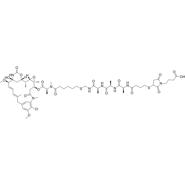 DM21-L-G
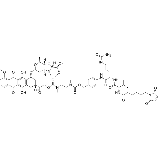 Mal-VC-PAB-DMEA-PNU-159682
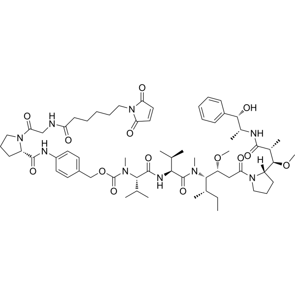 Mc-Pro-PAB-MMAE
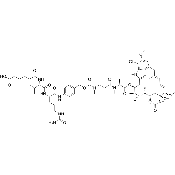 Maytansine derivative M24
InvivoChem的所有产品仅用于作科学研究,不面向患者销售
Copyright 2020 InvivoChem LLC | All Rights Reserved 粤ICP备20063088号-1


 COA
COA
