
| 规格 | 价格 | 库存 | 数量 |
|---|---|---|---|
| 1mg |
|
||
| 2mg |
|
||
| 5mg |
|
||
| 10mg |
|
||
| 25mg |
|
||
| 50mg |
|
||
| 100mg |
|
||
| Other Sizes |
|

| 靶点 |
Human Endogenous Metabolite; VDR/vitamin D receptor
|
||
|---|---|---|---|
| 体外研究 (In Vitro) |
Calcitriol 是 PHA 诱导的淋巴细胞增殖的有效抑制剂,培养 72 小时后可抑制 70% 的氚化胸苷掺入。骨化三醇以浓度依赖性方式减少 PHA 刺激的外周血单核细胞产生白细胞介素 2 (IL-2)。 [1]骨化三醇通过引起内质网释放钙并形成肌醇 1,4, 5-三磷酸和二酰基甘油,在不到 5 秒的时间内增加细胞内钙浓度 ([Ca2+]i)。 [2]骨化三醇既可以刺激又可以防止人类前列腺腺癌细胞的生长。骨化三醇可选择性降低 IV 型胶原酶(MMP-2 和 MMP-9)的分泌水平。 [3]骨化三醇可增加铂类药物的抗肿瘤活性,并在前列腺腺癌和鳞状细胞癌中表现出抗增殖活性。在 PC-3 和鼠鳞状细胞癌细胞中,与单独使用任一药物相比,在紫杉醇之前使用骨化三醇可显着降低克隆存活率。 [4]骨化三醇是一种有效的抗增殖剂,针对多种癌细胞类型。骨化三醇可调节生长因子受体表达,诱导细胞凋亡和分化,并增加 G0/G1 期停滞。骨化三醇抑制肿瘤细胞的运动和侵袭以及新血管的发育,从而放大多种细胞毒性药物的抗肿瘤作用。 [5]
|
||
| 体内研究 (In Vivo) |
骨化三醇治疗(每天 150 ng/kg,持续 4.5 个月)改善松弛(在用骨化三醇治疗的 OVX 中,pD2:6.30±0.09,Emax:68.6±3.9%,n=8)。 OVX 大鼠的两个肾脏的肾血流量均减少,可通过骨化三醇治疗来缓解。长期服用骨化三醇可降低 OVX 大鼠肾动脉中血栓素-前列腺素 (TP) 受体和 COX-2 的表达增加[3]。第 56 天,高剂量和低剂量骨化三醇治疗使果糖喂养大鼠的收缩压 (SBP) 分别降低 14±4 和 9±4 mmHg。与其他组相比,高剂量骨化三醇治疗(每天 20 ng/kg)显着升高血清离子钙水平(1.44±0.05 mmol/L)。
|
||
| 酶活实验 |
最近的研究表明,维生素D除了在维持钙稳态中具有明显的作用外,还可能具有其他重要的生物活性。在人外周血单核细胞和凝集素刺激的淋巴细胞中发现维生素D的胞浆受体,促使我们研究维生素D最具生物活性的代谢产物1,25-二羟基维生素D3(骨化三醇)对植物血凝素(PHA)诱导的淋巴细胞母细胞转化的影响。我们发现骨化三醇是PHA诱导的淋巴细胞增殖的有效抑制剂,在培养72小时后对氚化胸苷的掺入达到70%的抑制。此外,骨化三醇以浓度依赖的方式抑制PHA刺激的外周血单核细胞产生白细胞介素2(IL-2)。最后,通过添加饱和量的纯化IL-2,骨化三醇对细胞增殖的抑制作用被部分逆转。我们的结论是,骨化三醇是PHA诱导的淋巴细胞母细胞转化的有效抑制剂,这种作用在一定程度上是通过抑制IL-2的产生来介导的。因此,骨化三醇似乎具有迄今为止未被重视的免疫调节特性[1]。
|
||
| 细胞实验 |
骨化三醇 (100 nM) 或 DMSO(载体对照)用于治疗 CLL 细胞。在指定的时间间隔,通过轻度胰蛋白酶消化收获细胞。
通过克隆形成试验、3-(4,5-二甲基噻唑-2-基)-2,5-二苯基四唑溴化试验和监测肿瘤生长来测定治疗对小鼠鳞状细胞癌(SCCVII/SF)和人前列腺癌(PC-3)生长的影响。在紫杉醇之前用骨化三醇在体外处理SCC或PC-3细胞,与单独使用这两种药物相比,显著降低了克隆发生存活率。中剂量效应分析显示,骨化三醇和紫杉醇协同作用[4]。 |
||
| 动物实验 |
|
||
| 药代性质 (ADME/PK) |
Absorption, Distribution and Excretion
Upon administration, calcitriol is rapidly absorbed from the intestines. When a single oral dose of 0.5 mcg of calcitriol was administered, the mean serum concentrations of calcitriol rose from a baseline value of 40.0±4.4 (SD) pg/mL to 60.0±4.4 pg/mL at 2 hours, and declined to 53.0±6.9 at 4 hours, 50±7.0 at 8 hours, 44±4.6 at 12 hours and 41.5±5.1 at 24 hours. Following administration of single doses of 0.25 to 1.0 mcg of calcitriol, the peak plasma concentrations were reached within 3 to 6 hours. In a pharmacokinetic study, the oral bioavailability was 70.6±5.8% in healthy male volunteers and 72.2±4.8% in male patients with uraemia. In normal subjects, approximately 27% and 7% of the radioactivity appeared in the feces and urine, respectively, within 24 hours. Calcitriol undergoes enterohepatic recycling and biliary excretion. The metabolites of calcitriol are excreted primarily in feces. Cumulative excretion of radioactivity on the sixth day following intravenous administration of radiolabeled calcitriol averaged 16% in urine and 49% in feces. Upon intravenous administration, the volume of distribution of calcitriol was 0.49±0.14 L/kg in healthy male volunteers and 0.27±0.06 l/kg in uraemic male patients participating in a pharmacokinetic study. There is some evidence that calcitriol is transferred into human milk at low levels (ie, 2.2±0.1 pg/mL) in mothers. Calcitriol from maternal circulation may also enter the fetal circulation. The metabolic clearance rate was 23.5±4.34 ml/min in healthy male volunteers and 10.1±1.35 ml/min in male patients with uraemia. In the pediatric patients undergoing peritoneal dialysis receiving dose of 10.2 ng/kg (SD 5.5 ng/kg) for 2 months, the clearance rate was 15.3 mL/hr/kg. Many vitamin D analogs are readily absorbed from the GI tract following oral administration if fat absorption is normal. The presence of bile is required for absorption of ergocalciferol and the extent of GI absorption may be decreased in patients with hepatic, biliary, or GI disease (e.g., Crohn's disease, Whipple's disease, sprue). Because vitamin D is fat soluble, it is incorporated into chylomicrons and absorbed via the lymphatic system; approximately 80% of ingested vitamin D appears to be absorbed systemically through this mechanism, principally in the small intestine. Although some evidence suggested that intestinal absorption of vitamin D may be decreased in geriatric adults, other evidence did not show clinically important age-related alterations in GI absorption of the vitamin in therapeutic doses. It currently is not known whether aging alters the GI absorption of physiologic amounts of vitamin D. /Vitamin D analogs/ After oral administration of calcitriol, there is about a 2-hour lag-time before calcium absorption in the GI tract increases. Maximal hypercalcemic effect occurs in about 10 hours, and the duration of action of calcitriol is 3-5 days. Time to peak serum concentration: Oral: Approximately 3 to 6 hours. The primary route of excretion of vitamin D is the bile; only a small percentage of an administered dose is found in urine. /Vitamin D/ For more Absorption, Distribution and Excretion (Complete) data for 1,25-DIHYDROXYCHOLECALCIFEROL (10 total), please visit the HSDB record page. Metabolism / Metabolites Metabolism of calcitriol involves two pathways. The first pathway involves 24-hydroxylase activity in the kidney; this enzyme is also present in many target tissues which possess the vitamin D receptor such as the intestine. The end product of this pathway is a side chain shortened metabolite, calcitroic acid. The second pathway involves the conversion of calcitriol via the stepwise hydroxylation of carbon-26 and carbon-23, and cyclization to yield ultimately 1a,25R(OH)2-26,23S-lactone D3, which appears to be the major metabolite circulating in humans. Ohter identified metabolites of calcitriol include 1α, 25(OH)2-24-oxo-D3; 1α, 23,25(OH)3-24-oxo-D3; 1α, 24R,25(OH)3D3; 1α, 25S,26(OH)3D3; 1α, 25(OH)2-23-oxo-D3; 1α, 25R,26(OH)3-23-oxo-D3 and 1α, (OH)24,25,26,27-tetranor-COOH-D3. Calcitriol is the active form of vitamin D3 (cholecalciferol). The natural or endogenous supply of vitamin D in man mainly depends on ultraviolet light for conversion of 7-dehydrocholesterol to vitamin D3 in the skin. Vitamin D3 must be metabolically activated in the liver and the kidney before it is fully active on its target tissues. The initial transformation is catalyzed by a vitamin D3-25-hydroxylase enzyme present in the liver, and the product of this reaction is 25-(OH)D3 (calcifediol). The latter undergoes hydroxylation in the mitochondria of kidney tissue, and this reaction is activated by the renal 25-hydroxyvitamin D3-1-a-hydroxylase to produce 1,25-(OH)2D3 (calcitriol), the active form of vitamin D3. 1,25-Dihydroxycholecalciferol (calcitriol) and 1,25-dihydroxyergocalciferol appear to be metabolized to their respective trihydroxy metabolites (i.e., 1,24,25-trihydroxycholecalciferol, 1,24,25-trihydroxyergocalciferol) and to other compounds. The principal metabolite excreted in urine is calcitroic acid, which is more water soluble. Although all the metabolites of cholecalciferol and ergocalciferol have not been identified, hepatic microsomal enzymes may be involved in degrading metabolites of ergocalciferol and cholecalciferol. Calcitriol /(1,25-dihydroxy-vitamin D)/ is hydroxylated to 1,24,25-(OH)3-D by a renal hydroxylase that is induced by calcitriol and suppressed by those factors that stimulate the 25-OHD-1-alpha-hydroxylase. This enzyme also hydroxylates 25-OHD to form 24,25-(OH)2D. Both 24-hydroxylated compounds are less active than calcitriol and presumably represent metabolites destined for excretion. Side chain oxidation of calcitriol also occurs. To evaluate the relation between daily and fasting urinary calcium excretion and serum 1,25-dihydroxyvitamin D (II) concentrations, 6 healthy men were studied during control and during chronic oral calcitrol (I) administration (0.6, 1.2, or 1.8 nmols every 6 hours for 6-12 days) while they ate normal and low calcium diets (19.2 or 4.2 mmols Ca/day). Daily urinary calcium excretion was directly related to serum II concentrations, but increased more while subjects ate the normal calcium diet than when eating the low calcium diet. During I and ingestion of the low calcium diet, daily urinary calcium excretion averaged 7.32 mmole/day, exceeding the dietary calcium intake. Fasting urinary calcium/creatinine exceeded 0.34 mmol/mmol (the upper limit of normal) on either diet. When serum II concentrations are elevated, a high fasting urinary calcium/creatinine or high daily urinary calcium excretion, even on a low calcium diet, is insufficient criteria for the documentation of a renal calcium leak. For more Metabolism/Metabolites (Complete) data for 1,25-DIHYDROXYCHOLECALCIFEROL (7 total), please visit the HSDB record page. The first pathway involves 24-hydroxylase activity in the kidney; this enzyme is also present in many target tissues which possess the vitamin D receptor such as the intestine. The end product of this pathway is a side chain shortened metabolite, calcitroic acid. The second pathway involves the conversion of calcitriol via the stepwise hydroxylation of carbon-26 and carbon-23, and cyclization to yield ultimately 1a,25R(OH)2-26,23S-lactone D3. The lactone appears to be the major metabolite circulating in humans. Route of Elimination: Enterohepatic recycling and biliary excretion of calcitriol occur. The metabolites of calcitriol are excreted primarily in feces. Cumulative excretion of radioactivity on the sixth day following intravenous administration of radiolabeled calcitriol averaged 16% in urine and 49% in feces. Half Life: 5-8 hours Biological Half-Life After administration of single oral doses, the elimination half life was 5-8 hours. Plasma half-life: 3 to 6 hours. |
||
| 毒性/毒理 (Toxicokinetics/TK) |
Toxicity Summary
The mechanism of action of calcitriol in the treatment of psoriasis is accounted for by their antiproliferative activity for keratinocytes and their stimulation of epidermal cell differentiation. The anticarcinogenic activity of the active form of Calcitriol appears to be correlated with cellular vitamin D receptor (VDR) levels. Vitamin D receptors belong to the superfamily of steroid-hormone zinc-finger receptors. VDRs selectively bind 1,25-(OH)2-D3 and retinoic acid X receptor (RXR) to form a heterodimeric complex that interacts with specific DNA sequences known as vitamin D-responsive elements. VDRs are ligand-activated transcription factors. The receptors activate or repress the transcription of target genes upon binding their respective ligands. It is thought that the anticarcinogenic effect of Calcitriol is mediated via VDRs in cancer cells. The immunomodulatory activity of calcitriol is thought to be mediated by vitamin D receptors (VDRs) which are expressed constitutively in monocytes but induced upon activation of T and B lymphocytes. 1,25-(OH)2-D3 has also been found to enhance the activity of some vitamin D-receptor positive immune cells and to enhance the sensitivity of certain target cells to various cytokines secreted by immune cells. Effects During Pregnancy and Lactation ◉ Summary of Use during Lactation Calcitriol is the normal physiologically active form of vitamin D, 1,25-dihydroxyvitamin D. Several women with hypocalcemia have successfully breastfed during breastfeeding, with sometimes fluctuating serum calcium levels. Limited data indicate that its use in nursing mothers in appropriately adjusted doses does not affect the breastfed infant. If the mother requires calcitriol, it is not a reason to discontinue breastfeeding. Calcitriol and calcium dosage requirements are usually reduced during lactation in women with hypoparathyroidism. ◉ Effects in Breastfed Infants A woman with hypoparathyroidism breastfed her infant from week 1 to week 32 postpartum while taking calcitriol. The dose was initially 0.5 mcg daily, but was decreased to 0.25 mcg daily after 8 weeks. The infant thrived during breastfeeding and had normal serum calcium levels at 1 and 3 weeks and 3 months of age. A woman breastfed infants after two pregnancies while taking calcitriol in doses of 0.75 and 1 mcg daily. There were no reports of adverse reactions. A woman breastfed her newborn infant for 9 days while taking calcitriol 0.5 mcg three times daily. Calcitriol was stopped at that time because of hypercalcemia, but restarted at 40 days postpartum in low doses that were gradually increased until the prepregnancy dosage of 1.5 mcg daily was reached just before weaning at 12.5 months postpartum. A woman with discoid lupus was taking calcitriol 0.25 mcg every 2 days and several other medications concurrently. Her infant was breastfed for 12 months and followed up at 15 months of age. No adverse effects were reported during breastfeeding and the infant was growing and developing normally at 15 months of age. A nursing mother with autosomal dominant hypoparathyroidism type 1 was treated with teriparatide for 8 months postpartum then calcitriol 0.5 mcg twice daily was substituted. She breastfed her infant exclusively for 6 months then with supplementation to 1 year. Her infant had no change in serum calcium when maternal calcitriol was begun. The mother began weaning at 11 months and at 1 year of age weaning was complete. Growth and development were normal at 1.5 years of age. ◉ Effects on Lactation and Breastmilk Relevant published information was not found as of the revision date. Protein Binding Calcitriol is approximately 99.9% bound in blood, mostly by an alpha-globulin vitamin D binding protein. Toxicity Data LD50 (oral, rat) = 620 μg/kg; LD50 (intraperitoneal, rat) > 5 mg/kg. Interactions Corticosteroids counteract the effects of vitamin D analogs. /Vitamin D analogs/ Concurrent administration of thiazide diuretics and pharmacologic doses of vitamin D analogs in patients with hypoparathyroidism may result in hypercalcemia which may be transient and self-limited or may require discontinuance of vitamin D analogs. Thiazide-induced hypercalcemia in hypoparathyroid patients is probably caused by increased release of calcium from bone. /Vitamin D analogs/ Excessive use of mineral oil may interfere with intestinal absorption of vitamin D analogs. /Vitamin D analogs/ Orlistat may result in decreased GI absorption of fat-soluble vitamins such as vitamin D analogs. At least 2 hours should elapse between (before or after) any orlistat dose and vitamin D analog administration ... . /Vitamin D analogs/ For more Interactions (Complete) data for 1,25-DIHYDROXYCHOLECALCIFEROL (8 total), please visit the HSDB record page. |
||
| 参考文献 | |||
| 其他信息 |
Therapeutic Uses
Bone Density Conservation Agents; Calcium Channel Agonist; Vitamins Calcium Channel Agonists; Dermatologic Agents Medication: Calcium regulator; vitamin (antirachitic) Therapeutic doses of specific vitamin D analogs are used in the treatment of chronic hypocalcemia, hypophosphatemia, rickets, and osteodystrophy associated with various medical conditions including chronic renal failure, familial hypophosphatemia, and hypoparathyroidism (postsurgical or idiopathic, or pseudohypoparathyroidism). Some analogs have been found to reduct elevated parathyroid hormone concentrations in patients with renal osteodystrophy associated with hyperparathyroidism. Theoretically, any of the vitamin D analogs may be used for the above conditions, However, because of their pharmacologic properties, some may be more useful in certain situations than others. Alfacalcidol, calcitriol, and dihydrotachysterol are usually preferred in patients with renal failure since these patients have impaired ability to synthesize calcitriol from cholecalciferol and ergocalciferol; therefore, the response is more predictable. In addition, their shorter half-lives may make toxicity easier to manage (hypercalcemia reverses more quickly). Ergocalciferol may not be the preferred agent in the treatment of familial hypophosphatemia or hypoparathyroidism because the large doses needed are associated with a risk of overdose and hypercalcemia; dihydrotachysterol and calcitriol may be preferred. /Included in US product labeling/ For more Therapeutic Uses (Complete) data for 1,25-DIHYDROXYCHOLECALCIFEROL (6 total), please visit the HSDB record page. Drug Warnings ... When serum alkaline phosphate decreases, serum calcium rises. Metastatic calcification, decrease in renal function, and increased serum phosphate levels are possible consequences. POTENTIAL ADVERSE EFFECTS ON FETUS: Teratogenic in animals at high doses (4-15x recommended human dose). In humans, maternal hypercalcemia during pregnancy may increase fetal sensitivity to effects of vitamin D, suppression of parathyroid function or a syndrome of elfin facies, mental retardation, and congenital supravalvular aortic stenosis. POTENTIAL SIDE EFFECTS ON BREAST-FED INFANT: No known problems at recommended daily allowance. /Cholecalciferol from table II/ Doses of vitamin D analogs that do not exceed the physiologic requirement are usually nontoxic. However, some infants and patients with sarcoidosis or hypoparathyroidism may have increased sensitivity to vitamin D analogs. /Vitamin D analogs/ Decreased renal function without hypercalcemia has also been reported in patients with hypoparathyroidism after long-term vitamin D analog therapy. Before therapy with vitamin D analogs is initiated, serum phosphate concentrations must be controlled. To avoid ectopic calcification, the serum calcium (in mg/dL) times phosphorus (in mg/dL) should not be allowed to exceed 70. Because administration of vitamin D analogs may increase phosphate absorption, patients with renal failure may require adjustment in the dosage of aluminum-containing antacids used to decrease phosphate absorption. /Vitamin D analogs/ For more Drug Warnings (Complete) data for 1,25-DIHYDROXYCHOLECALCIFEROL (13 total), please visit the HSDB record page. Pharmacodynamics Calcitriol is a biologically active calcitrophic hormone with anti-osteoporotic, immunomodulatory, anticarcinogenic, antipsoriatic, antioxidant, and mood-modulatory activities. Its main sites of action are the intestine, bone, kidney and parathyroid hormone. Calcitriol is a ligand for the vitamin D nuclear receptor, which is expressed in, but not limited to, gastrointestinal (GI) tissues, bones, and kidneys. As an active form of vitamin D3, calcitriol elevates the plasma levels of calcium by stimulating intestinal calcium uptake, increasing reabsorption of calcium by the kidneys, and possibly increasing the release of calcium from skeletal stores. The duration of pharmacologic activity of a single dose of exogenous calcitriol is expected to be about 3 to 5 days. In addition to its important role in calcium metabolism, other pharmacological effects of calcitriol have been studied in various conditions including cancer models. Various studies demonstrated expression of vitamin D receptors in cancer cell lines, including mouse myeloid leukemia cells. Calcitriol has been found to induce differentiation and/or inhibit cell proliferation _in vitro_ and _in vivo_ in many cell types, such as malignant cell lines carcinomas of the breast, prostate, colon, skin, and brain, myeloid leukemia cells, and others. In early human prostate cancer trials, administration of 1.5 µg/d calcitriol in male participants resulted in a reduction in the rate of PSA rise in most participants, however it was coincided with dose-limiting hypercalcemia in most participants. Hypercalcemia and hypercalcuria were evident in numerous initial trials, and this may be due to these trials not testing the drug at concentrations that are active in preclinical systems. Findings from preclinical data show an additive or synergistic antineoplastic action of calcitriol when combined with agents including dexamethasone, retinoids, and radiation, as well as several cytotoxic chemotherapy drugs such as platinum compounds. Vitamin D deficiency has long been suspected to increase the susceptibility to tuberculosis. The active form of calcitriol, 1,25-(OH)2-D3, has been found to enhance the ability of mononuclear phagocytes to suppress the intracellular growth of Mycobacterium tuberculosis. 1,25-(OH)2-D3 has demonstrated beneficial effects in animal models of such autoimmune diseases as rheumatoid arthritis. Vitamin D appears to demonstrate both immune-enhancing and immunosuppressive effects. |
| 分子式 |
C27H44O3
|
|
|---|---|---|
| 分子量 |
416.64
|
|
| 精确质量 |
416.329
|
|
| 元素分析 |
C, 77.84; H, 10.65; O, 11.52
|
|
| CAS号 |
32222-06-3
|
|
| 相关CAS号 |
(1S)-Calcitriol;61476-45-7;Calcitriol-d6;78782-99-7;Calcitriol-13C3;Calcitriol-d3;128723-16-0;Calcitriol Derivatives;2070009-24-2
|
|
| PubChem CID |
5280453
|
|
| 外观&性状 |
White to off-white solid powder
|
|
| 密度 |
1.1±0.1 g/cm3
|
|
| 沸点 |
565.0±50.0 °C at 760 mmHg
|
|
| 熔点 |
119-1210C
|
|
| 闪点 |
238.4±24.7 °C
|
|
| 蒸汽压 |
0.0±3.5 mmHg at 25°C
|
|
| 折射率 |
1.547
|
|
| LogP |
6.12
|
|
| tPSA |
60.69
|
|
| 氢键供体(HBD)数目 |
3
|
|
| 氢键受体(HBA)数目 |
3
|
|
| 可旋转键数目(RBC) |
6
|
|
| 重原子数目 |
30
|
|
| 分子复杂度/Complexity |
688
|
|
| 定义原子立体中心数目 |
6
|
|
| SMILES |
C=C1[C@H](C[C@@H](C/C1=C/C=C2[C@]3([C@@](C)([C@H](CC3)[C@@H](CCCC(C)(O)C)C)CCC/2)[H])O)O
|
|
| InChi Key |
GMRQFYUYWCNGIN-NKMMMXOESA-N
|
|
| InChi Code |
InChI=1S/C27H44O3/c1-18(8-6-14-26(3,4)30)23-12-13-24-20(9-7-15-27(23,24)5)10-11-21-16-22(28)17-25(29)19(21)2/h10-11,18,22-25,28-30H,2,6-9,12-17H2,1,3-5H3/b20-10+,21-11-/t18-,22-,23-,24+,25+,27-/m1/s1
|
|
| 化学名 |
(1R,3S,5Z)-5-[(2E)-2-[(1R,3aS,7aR)-1-[(2R)-6-hydroxy-6-methylheptan-2-yl]-7a-methyl-2,3,3a,5,6,7-hexahydro-1H-inden-4-ylidene]ethylidene]-4-methylidenecyclohexane-1,3-diol
|
|
| 别名 |
|
|
| HS Tariff Code |
2934.99.9001
|
|
| 存储方式 |
Powder -20°C 3 years 4°C 2 years In solvent -80°C 6 months -20°C 1 month 注意: (1). 本产品在运输和储存过程中需避光。 (2). 请将本产品存放在密封且受保护的环境中(例如氮气保护),避免吸湿/受潮。 (3). 该产品在溶液状态不稳定,请现配现用。 |
|
| 运输条件 |
Room temperature (This product is stable at ambient temperature for a few days during ordinary shipping and time spent in Customs)
|
| 溶解度 (体外实验) |
|
|||
|---|---|---|---|---|
| 溶解度 (体内实验) |
配方 1 中的溶解度: 2.75 mg/mL (6.60 mM) in 10% DMSO + 40% PEG300 + 5% Tween80 + 45% Saline (这些助溶剂从左到右依次添加,逐一添加), 悬浮液;超声助溶。
例如,若需制备1 mL的工作液,可将100 μL 27.5 mg/mL澄清DMSO储备液加入400 μL PEG300中,混匀;然后向上述溶液中加入50 μL Tween-80,混匀;加入450 μL生理盐水定容至1 mL。 *生理盐水的制备:将 0.9 g 氯化钠溶解在 100 mL ddH₂O中,得到澄清溶液。 配方 2 中的溶解度: ≥ 2.75 mg/mL (6.60 mM) (饱和度未知) in 10% DMSO + 90% (20% SBE-β-CD in Saline) (这些助溶剂从左到右依次添加,逐一添加), 澄清溶液。 例如,若需制备1 mL的工作液,可将 100 μL 27.5mg/mL澄清的DMSO储备液加入到900μL 20%SBE-β-CD生理盐水中,混匀。 *20% SBE-β-CD 生理盐水溶液的制备(4°C,1 周):将 2 g SBE-β-CD 溶解于 10 mL 生理盐水中,得到澄清溶液。 View More
配方 3 中的溶解度: ≥ 2.75 mg/mL (6.60 mM) (饱和度未知) in 10% DMSO + 90% Corn Oil (这些助溶剂从左到右依次添加,逐一添加), 澄清溶液。 配方 4 中的溶解度: ≥ 2.75 mg/mL (6.60 mM) (饱和度未知) in 5% DMSO + 40% PEG300 + 5% Tween80 + 50% Saline (这些助溶剂从左到右依次添加,逐一添加), 澄清溶液。 *生理盐水的制备:将 0.9 g 氯化钠溶解在 100 mL ddH₂O中,得到澄清溶液。 配方 5 中的溶解度: ≥ 2.75 mg/mL (6.60 mM) (饱和度未知) in 5% DMSO + 95% (20% SBE-β-CD in Saline) (这些助溶剂从左到右依次添加,逐一添加), 澄清溶液。 *20% SBE-β-CD 生理盐水溶液的制备(4°C,1 周):将 2 g SBE-β-CD 溶解于 10 mL 生理盐水中,得到澄清溶液。 配方 6 中的溶解度: ≥ 2.5 mg/mL (6.00 mM) (饱和度未知) in 10% EtOH + 40% PEG300 + 5% Tween80 + 45% Saline (这些助溶剂从左到右依次添加,逐一添加), 澄清溶液。 例如,若需制备1 mL的工作液,将 100 μL 25.0 mg/mL 澄清乙醇储备液加入到 400 μL PEG300 中,混匀;然后向上述溶液中加入50 μL Tween-80,混匀;加入450 μL生理盐水定容至1 mL。 *生理盐水的制备:将 0.9 g 氯化钠溶解在 100 mL ddH₂O中,得到澄清溶液。 配方 7 中的溶解度: ≥ 2.5 mg/mL (6.00 mM) (饱和度未知) in 10% EtOH + 90% (20% SBE-β-CD in Saline) (这些助溶剂从左到右依次添加,逐一添加), 澄清溶液。 例如,要配制1 mL工作液,可将100 μL 25.0 mg/mL的澄清EtOH储备液加入到900 μL 20% SBE-β-CD生理盐水溶液中, 混合均匀。 *20% SBE-β-CD 生理盐水溶液的制备(4°C,1 周):将 2 g SBE-β-CD 溶解于 10 mL 生理盐水中,得到澄清溶液。 配方 8 中的溶解度: ≥ 2.5 mg/mL (6.00 mM) (饱和度未知) in 10% EtOH + 90% Corn Oil (这些助溶剂从左到右依次添加,逐一添加), 澄清溶液。 例如,要配制1 mL工作液,可将100 μL 25.0 mg/mL 澄清乙醇储备液加入到900 μL 玉米油中,混匀。 配方 9 中的溶解度: 0.55 mg/mL (1.32 mM) in 1% DMSO 99% Saline (这些助溶剂从左到右依次添加,逐一添加), 悬浊液; 超声助溶。 *生理盐水的制备:将 0.9 g 氯化钠溶解在 100 mL ddH₂O中,得到澄清溶液。 1、请先配制澄清的储备液(如:用DMSO配置50 或 100 mg/mL母液(储备液)); 2、取适量母液,按从左到右的顺序依次添加助溶剂,澄清后再加入下一助溶剂。以 下列配方为例说明 (注意此配方只用于说明,并不一定代表此产品 的实际溶解配方): 10% DMSO → 40% PEG300 → 5% Tween-80 → 45% ddH2O (或 saline); 假设最终工作液的体积为 1 mL, 浓度为5 mg/mL: 取 100 μL 50 mg/mL 的澄清 DMSO 储备液加到 400 μL PEG300 中,混合均匀/澄清;向上述体系中加入50 μL Tween-80,混合均匀/澄清;然后继续加入450 μL ddH2O (或 saline)定容至 1 mL; 3、溶剂前显示的百分比是指该溶剂在最终溶液/工作液中的体积所占比例; 4、 如产品在配制过程中出现沉淀/析出,可通过加热(≤50℃)或超声的方式助溶; 5、为保证最佳实验结果,工作液请现配现用! 6、如不确定怎么将母液配置成体内动物实验的工作液,请查看说明书或联系我们; 7、 以上所有助溶剂都可在 Invivochem.cn网站购买。 |
| 制备储备液 | 1 mg | 5 mg | 10 mg | |
| 1 mM | 2.4002 mL | 12.0008 mL | 24.0015 mL | |
| 5 mM | 0.4800 mL | 2.4002 mL | 4.8003 mL | |
| 10 mM | 0.2400 mL | 1.2001 mL | 2.4002 mL |
1、根据实验需要选择合适的溶剂配制储备液 (母液):对于大多数产品,InvivoChem推荐用DMSO配置母液 (比如:5、10、20mM或者10、20、50 mg/mL浓度),个别水溶性高的产品可直接溶于水。产品在DMSO 、水或其他溶剂中的具体溶解度详见上”溶解度 (体外)”部分;
2、如果您找不到您想要的溶解度信息,或者很难将产品溶解在溶液中,请联系我们;
3、建议使用下列计算器进行相关计算(摩尔浓度计算器、稀释计算器、分子量计算器、重组计算器等);
4、母液配好之后,将其分装到常规用量,并储存在-20°C或-80°C,尽量减少反复冻融循环。
计算结果:
工作液浓度: mg/mL;
DMSO母液配制方法: mg 药物溶于 μL DMSO溶液(母液浓度 mg/mL)。如该浓度超过该批次药物DMSO溶解度,请首先与我们联系。
体内配方配制方法:取 μL DMSO母液,加入 μL PEG300,混匀澄清后加入μL Tween 80,混匀澄清后加入 μL ddH2O,混匀澄清。
(1) 请确保溶液澄清之后,再加入下一种溶剂 (助溶剂) 。可利用涡旋、超声或水浴加热等方法助溶;
(2) 一定要按顺序加入溶剂 (助溶剂) 。
Vitamin D as a Therapeutic Adjunct in the Stimulant Treatment of ADHD
CTID: NCT03103750
Phase: Phase 1 Status: Completed
Date: 2024-03-19
 Calcipotriol inducedERαexpression in ER-negative breast cancer cells.
Calcitriol induced ERαprotein expression.BMC Cancer.2014 Mar 29;14:230. doi: 10.1186/1471-2407-14-230. |
|---|
 Immunocytochemical analysis of ERαand VDR in primary and established breast cancer cells.BMC Cancer.2014 Mar 29;14:230. doi: 10.1186/1471-2407-14-230.
Calcitriol induced a fully active ERα.BMC Cancer.2014 Mar 29;14:230. doi: 10.1186/1471-2407-14-230. |
 Calcitriol inducedERαmRNA expression through the VDR in ERα-negative breast cancer cells.BMC Cancer.2014 Mar 29;14:230. doi: 10.1186/1471-2407-14-230. |
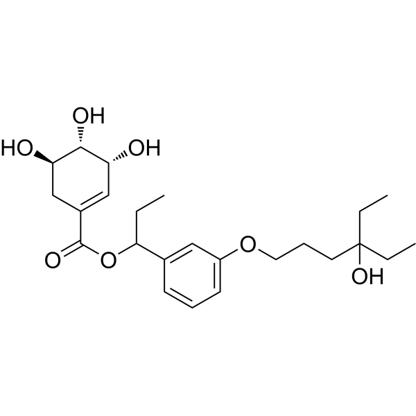 VDR agonist 3
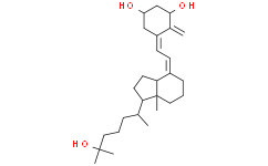 (1R,5Z)-Calcitriol
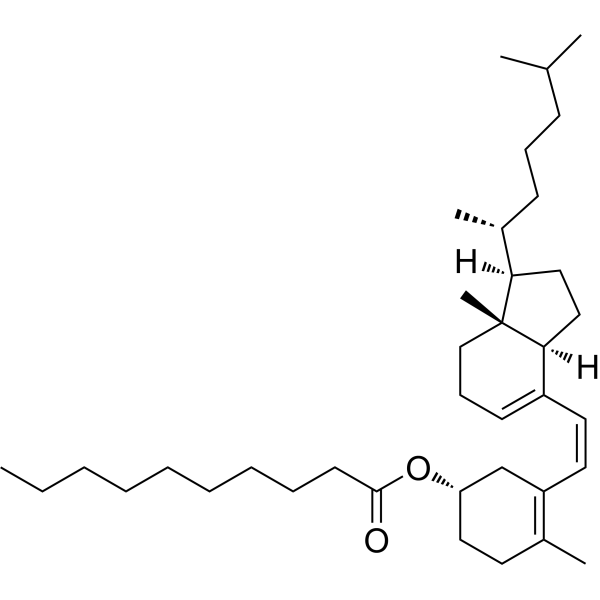 Pre-vitamin D3 decanoate
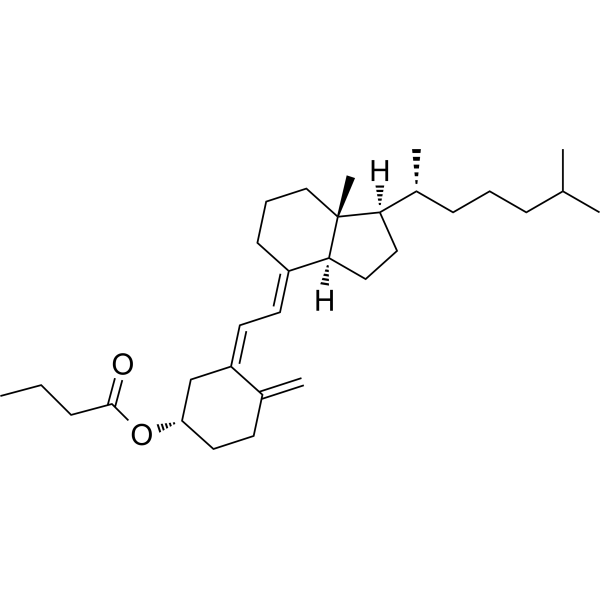 Butyrate-Vitamin D3
InvivoChem的所有产品仅用于作科学研究,不面向患者销售
Copyright 2020 InvivoChem LLC | All Rights Reserved 粤ICP备20063088号-1


 COA
COA


