
| 规格 | 价格 | 库存 | 数量 |
|---|---|---|---|
| 10 mM * 1 mL in DMSO |
|
||
| 1mg |
|
||
| 5mg |
|
||
| 10mg |
|
||
| 25mg |
|
||
| 50mg |
|
||
| 100mg |
|
||
| 250mg |
|
||
| Other Sizes |
|

| 靶点 |
Activated mTRESK channel (IC50 = 6.8 μM)
|
|---|---|
| 体外研究 (In Vitro) |
将 A2764 (100 µM) 应用于表达 mTRESK 的卵母细胞可导致背景 K+ 电流抑制 42.8±11.5%[1]。在离子霉素诱导的 mTRESK 电流中,A2764 (100 µM) 与激活通道的 IC50 相比表现出增强的抑制活性。之后,使用A2764显着降低电流(77.8±3.5%)[1]。在静息条件下,A2764 (100 µM) 可使 TRESK 电流降低 42.8±11.5%,而在活动期间,它会抑制 TRESK 电流 77.8±3.5% [1]。
|
| 酶活实验 |
双电极电压钳和膜片钳测量。[1]
如前所述,在将cRNA显微注射到爪蟾卵母细胞中1-3天后进行了双电极电压钳实验(Czirják等人,2004)。对于每种通道类型,对n个数有贡献的卵母细胞(确切的n个数在文本或图中标明)来自至少两个,但通常是三个单独的青蛙。保持电位为0 mV。在每4秒施加300毫秒长的电压阶跃至-100 mV后,测量背景钾电流。低钾记录溶液含有以下物质(单位为mM):NaCl 95.4、KCl 2、CaCl2 1.8和HEPES 5,pH值为7.5,用NaOH调节。高钾溶液含有80mM K+(低钾溶液中的78mM NaCl被KCl替代)。为了测量TREK-1、TREK-2和TRAAK电流,高钾溶液含有40 mM K+。使用重力驱动灌注系统将溶液施加到卵母细胞上。实验在室温(21°C)下进行 电压钳配置中的全细胞膜片钳实验如前所述进行(Lengyel等人,2016)。在无电流注入的电流钳模式下记录静息膜电位(I=0模式)。通过每4秒注入去极化电流(以100 pA为增量,高达1500 pA)1秒来测定流变碱。分离后1至2天,将分离的DRG神经元用于实验。对于DRG神经元的电流钳研究,如先前的研究所述,只接受膜电位在-45至-70 mV之间的细胞(Petruska等人,2000)。由于本研究的重点是检查我们新的氯氧喹类似物对分离的DRG神经元电生理参数的影响,因此没有使用其他选择标准。将八极点贝塞尔滤波器的截止频率调整为200Hz,并在1kHz下采集数据。移液管溶液含有(以mM计):140 KCl、3 MgCl2、0.05 EGTA、1 Na2 ATP、0.1 Na2 GTP和10 HEPES。低钾溶液含有(以mM计):140 NaCl、3.6 KCl、0.5 MgCl2、2 CaCl2、11葡萄糖和10 HEPES。高钾溶液含有30mM KCl(低钾溶液的26.4mM NaCl被KCl代替)。用NaOH将浴溶液的pH值调节至7.4。实验在室温(21°C)下进行。 |
| 动物实验 |
Animal Husbandry, Preparation, and Microinjection of Xenopus Oocytes. Generation of a TRESK Knockout Mouse Line: Isolation of DRG Neurons.[1]
Xenopus laevis oocytes were prepared as previously described (Czirják and Enyedi, 2002). For the expression of the different channels, the oocytes were injected with 57 pg to 4 ng of cRNA (depending on the channel type) 1 day after defolliculation. Injection was performed with a Nanoliter Injector. X. laevis frogs were housed in 50-l tanks with continuous filtering and water circulation. Room temperature was 19°C. Frogs were anesthetized with 0.1% tricaine solution and killed by decerebration and pithing. FVB/Ant (FVB.129P2-Pde6b+Tyrc-ch/Ant) mice were obtained commercially. TRESK knockout (KO) animals were generated by the transcription activator–like effector nuclease (TALEN) technique using plasmids ordered from Addgene. Mouse TRESK (mTRESK)-specific TALEN recognition sites were designed for the genomic sequence of the first exon (corresponding to the N-terminal intracellular domain of the channel) using the 5′-TN19 N14−20 N19A-3′ formula, with the following sequences: left TALEN recognition site, 5′tgaggagccacctgaggcca; right TALEN recognition site, 5′ccctggggaaggccagggga; and an 18 base pair (5′ggagatgctgtcctgagg) FokI nuclease dimerization and cutting sequence in between. mTRESK recognizing TALEN plasmids were assembled according to the protocol of Sanjana et al. (2012). TALEN mRNAs were produced using the Ambion mMESSAGE mMACHINE T7 in vitro transcription kit (Ambion). TALEN mRNAs were microinjected at a concentration of 20–20 ng/μl into the pronuclei of fertilized eggs of FVB/Ant mice. Pups were analyzed with Surveyor assay plus sequencing. In 12 mice, among the 61 born pups the TRESK (KCNK18) gene was changed, and a founder bearing a 33 base pair deletion and also a mutation introducing a stop codon was chosen to establish a colony. Adult female wild-type and TRESK KO mice (2–3 months of age) were used for the patch-clamp experiments in this study. The animals were maintained on a 12-hour light/dark cycle with free access to food and water in a specific pathogen-free animal facility. Mice were killed humanely by CO2 exposure (CO2 was applied until death of the animals). DRGs were dissected from the thoracic and lumbar levels of the spinal cord and collected in sterile PBS (137 mM NaCl, 2.7 mM KCl, and 10 mM NaH2PO4, pH adjusted to 7.4 with NaOH) at 4°C. Ganglia were incubated in PBS containing 2 mg/ml collagenase enzyme (type I) for 30 minutes with gentle shaking at 37°C. For further details regarding the isolation and culturing of the cells, see Braun et al. (2015). All experimental procedures using animals were conducted in accordance with the Guide for the Care and Use of Laboratory Animals as adopted by the National Institutes of Health, local state laws, and institutional regulations. All animal experiments were approved by the Animal Care and Ethics Committee of Semmelweis University (approval ID: XIV-I-001/2154-4/2012). |
| 参考文献 | |
| 其他信息 |
Cloxyquin has been reported as a specific activator of TRESK [TWIK-related spinal cord K+ channel (also known as K2P18.1)] background potassium channel. In this study, we have synthetized chemically modified analogs of cloxyquin and tested their effects on TRESK and other K2P channels. The currents of murine K2P channels, expressed heterologously in Xenopus oocytes, were measured by two-electrode voltage clamp, whereas the native background K+ conductance of mouse dorsal root ganglion (DRG) neurons was examined by the whole-cell patch-clamp method. Some of the analogs retained the activator character of the parent compound, but, more interestingly, other derivatives inhibited mouse TRESK current. The inhibitor analogs (A2764 and A2793) exerted state-dependent effects. The degree of inhibition by 100 µM A2764 (77.8% ± 3.5%, n = 6) was larger in the activated state of TRESK (i.e., after calcineurin-dependent stimulation) than in the resting state of the channel (42.8% ± 11.5% inhibition, n = 7). The selectivity of the inhibitor compounds was tested on several K2P channels. A2793 inhibited TWIK-related acid-sensitive K+ channel (TASK)-1 (100 µM, 53.4% ± 13, 5%, n = 5), while A2764 was more selective for TRESK, it only moderately influenced TREK-1 and TWIK-related alkaline pH-activated K+ channel. The effect of A2764 was also examined on the background K+ currents of DRG neurons. A subpopulation of DRG neurons, prepared from wild-type animals, expressed background K+ currents sensitive to A2764, whereas the inhibitor did not affect the currents in the DRG neurons of TRESK-deficient mice. Accordingly, A2764 may prove to be useful for the identification of TRESK current in native cells, and for the investigation of the role of the channel in nociception and migraine. SIGNIFICANCE STATEMENT: TRESK background potassium channel is a potential pharmacological target in migraine and neuropathic pain. In this study, we have identified a selective inhibitor of TRESK, A2764. This compound can inhibit TRESK in native cells, leading to cell depolarization and increased excitability. This new inhibitor may be of use to probe the role of TRESK channel in migraine and nociception.[1]
|
| 分子式 |
C15H20CL2N2O
|
|---|---|
| 分子量 |
315.238101959229
|
| 精确质量 |
350.071
|
| CAS号 |
861038-72-4
|
| PubChem CID |
146013302
|
| 外观&性状 |
Typically exists as Light yellow to yellow solids at room temperature
|
| tPSA |
25.4Ų
|
| 氢键供体(HBD)数目 |
2
|
| 氢键受体(HBA)数目 |
3
|
| 可旋转键数目(RBC) |
6
|
| 重原子数目 |
21
|
| 分子复杂度/Complexity |
261
|
| 定义原子立体中心数目 |
0
|
| InChi Key |
ZVRDPULCSHGKBD-UHFFFAOYSA-N
|
| InChi Code |
InChI=1S/C15H19ClN2O.2ClH/c1-3-18(4-2)10-11-19-14-8-7-13(16)12-6-5-9-17-15(12)14;;/h5-9H,3-4,10-11H2,1-2H3;2*1H
|
| 化学名 |
2-(5-chloroquinolin-8-yl)oxy-N,N-diethylethanamine;dihydrochloride
|
| 别名 |
A 2764 diHCl; A-2764 diHCl; A2764 dihydrochloride; 2-((5-Chloroquinolin-8-yl)oxy)-N,N-diethylethanamine dihydrochloride; A2764 (dihydrochloride); 2-((5-Chloroquinolin-8-yl)oxy)-N,N-diethylethanaminedihydrochloride; 2-(5-chloroquinolin-8-yl)oxy-N,N-diethylethanamine;dihydrochloride; A2764 dihydrochloride?; TRESK inhibitor A2764;
|
| HS Tariff Code |
2934.99.9001
|
| 存储方式 |
Powder -20°C 3 years 4°C 2 years In solvent -80°C 6 months -20°C 1 month 注意: 请将本产品存放在密封且受保护的环境中,避免吸湿/受潮。 |
| 运输条件 |
Room temperature (This product is stable at ambient temperature for a few days during ordinary shipping and time spent in Customs)
|
| 溶解度 (体外实验) |
H2O : ≥ 100 mg/mL (~284.33 mM)
DMSO : ~41.67 mg/mL (~118.48 mM) |
|---|---|
| 溶解度 (体内实验) |
配方 1 中的溶解度: ≥ 2.08 mg/mL (5.91 mM) (饱和度未知) in 10% DMSO + 40% PEG300 + 5% Tween80 + 45% Saline (这些助溶剂从左到右依次添加,逐一添加), 澄清溶液。
例如,若需制备1 mL的工作液,可将100 μL 20.8 mg/mL澄清DMSO储备液加入400 μL PEG300中,混匀;然后向上述溶液中加入50 μL Tween-80,混匀;加入450 μL生理盐水定容至1 mL。 *生理盐水的制备:将 0.9 g 氯化钠溶解在 100 mL ddH₂O中,得到澄清溶液。 配方 2 中的溶解度: ≥ 2.08 mg/mL (5.91 mM) (饱和度未知) in 10% DMSO + 90% (20% SBE-β-CD in Saline) (这些助溶剂从左到右依次添加,逐一添加), 澄清溶液。 例如,若需制备1 mL的工作液,可将 100 μL 20.8 mg/mL澄清DMSO储备液加入900 μL 20% SBE-β-CD生理盐水溶液中,混匀。 *20% SBE-β-CD 生理盐水溶液的制备(4°C,1 周):将 2 g SBE-β-CD 溶解于 10 mL 生理盐水中,得到澄清溶液。 请根据您的实验动物和给药方式选择适当的溶解配方/方案: 1、请先配制澄清的储备液(如:用DMSO配置50 或 100 mg/mL母液(储备液)); 2、取适量母液,按从左到右的顺序依次添加助溶剂,澄清后再加入下一助溶剂。以 下列配方为例说明 (注意此配方只用于说明,并不一定代表此产品 的实际溶解配方): 10% DMSO → 40% PEG300 → 5% Tween-80 → 45% ddH2O (或 saline); 假设最终工作液的体积为 1 mL, 浓度为5 mg/mL: 取 100 μL 50 mg/mL 的澄清 DMSO 储备液加到 400 μL PEG300 中,混合均匀/澄清;向上述体系中加入50 μL Tween-80,混合均匀/澄清;然后继续加入450 μL ddH2O (或 saline)定容至 1 mL; 3、溶剂前显示的百分比是指该溶剂在最终溶液/工作液中的体积所占比例; 4、 如产品在配制过程中出现沉淀/析出,可通过加热(≤50℃)或超声的方式助溶; 5、为保证最佳实验结果,工作液请现配现用! 6、如不确定怎么将母液配置成体内动物实验的工作液,请查看说明书或联系我们; 7、 以上所有助溶剂都可在 Invivochem.cn网站购买。 |
| 制备储备液 | 1 mg | 5 mg | 10 mg | |
| 1 mM | 3.1722 mL | 15.8609 mL | 31.7219 mL | |
| 5 mM | 0.6344 mL | 3.1722 mL | 6.3444 mL | |
| 10 mM | 0.3172 mL | 1.5861 mL | 3.1722 mL |
1、根据实验需要选择合适的溶剂配制储备液 (母液):对于大多数产品,InvivoChem推荐用DMSO配置母液 (比如:5、10、20mM或者10、20、50 mg/mL浓度),个别水溶性高的产品可直接溶于水。产品在DMSO 、水或其他溶剂中的具体溶解度详见上”溶解度 (体外)”部分;
2、如果您找不到您想要的溶解度信息,或者很难将产品溶解在溶液中,请联系我们;
3、建议使用下列计算器进行相关计算(摩尔浓度计算器、稀释计算器、分子量计算器、重组计算器等);
4、母液配好之后,将其分装到常规用量,并储存在-20°C或-80°C,尽量减少反复冻融循环。
计算结果:
工作液浓度: mg/mL;
DMSO母液配制方法: mg 药物溶于 μL DMSO溶液(母液浓度 mg/mL)。如该浓度超过该批次药物DMSO溶解度,请首先与我们联系。
体内配方配制方法:取 μL DMSO母液,加入 μL PEG300,混匀澄清后加入μL Tween 80,混匀澄清后加入 μL ddH2O,混匀澄清。
(1) 请确保溶液澄清之后,再加入下一种溶剂 (助溶剂) 。可利用涡旋、超声或水浴加热等方法助溶;
(2) 一定要按顺序加入溶剂 (助溶剂) 。
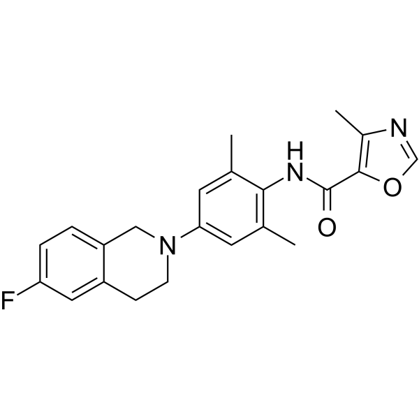 Kv7.2 modulator 1
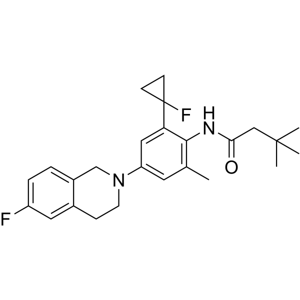 Kv7.2 modulator 2
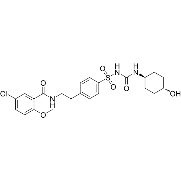 4-trans-Hydroxy-glibenclamide
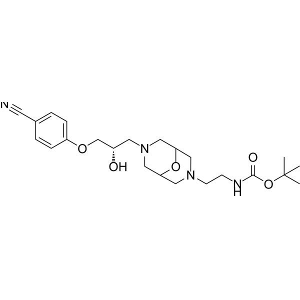 Inakalant
InvivoChem的所有产品仅用于作科学研究,不面向患者销售
Copyright 2020 InvivoChem LLC | All Rights Reserved 粤ICP备20063088号-1


 COA
COA
